

mTOR activation, lymphangiogenesis, and estrogen-mediated cell survival: the "perfect storm" of pro-metastatic factors in LAM pathogenesis
- PMID: 20235886
- PMCID: PMC2883473
- DOI: 10.1089/lrb.2009.0020
mTOR activation, lymphangiogenesis, and estrogen-mediated cell survival: the "perfect storm" of pro-metastatic factors in LAM pathogenesis
Abstract
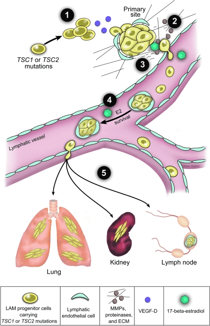
Research interest in lymphangioleiomyomatosis (LAM) has grown dramatically in the past decade, particularly among cancer biologists. There are at least two reasons for this: first, the discovery in the year 2000 that LAM cells carry TSC2 gene mutations, linking LAM with cellular pathways including the PI3K/Akt/mTOR axis, and allowing the Tuberous Sclerosis Complex (TSC)-regulated pathways that are believed to underlie LAM pathogenesis to be studied in cells, yeast, Drosophila, and mice. A second reason for the rising interest in LAM is the discovery that LAM cells can travel to the lung, including repopulating a donor lung after lung transplantation, despite the fact that LAM cells are histologically benign. This "benign metastasis" underpinning suggests that elucidating LAM pathogenesis will unlock a set of fundamental mechanisms that underlie metastatic potential in the context of a cell that has not yet undergone malignant transformation. Here, we will outline the data supporting the metastatic model of LAM, consider the biochemical and cellular mechanisms that may enable LAM cells to metastasize, including both cell autonomous and non-cell autonomous factors, and highlight a mouse model in which estrogen promotes the metastasis and survival of TSC2-deficient cells in a MEK-dependent manner. We propose a multistep model of LAM cell metastasis that highlights multiple opportunities for therapeutic intervention. Taken together, the metastatic behavior of LAM cells and the involvement of tumor-related signaling pathways lead to optimism that cancer-related paradigms for diagnosis, staging, and therapy will lead to therapeutic breakthroughs for women living with LAM.
Figures

Similar articles
-
Animal models of lymphangioleiomyomatosis (LAM) and tuberous sclerosis complex (TSC).Lymphat Res Biol. 2010 Mar;8(1):51-7. doi: 10.1089/lrb.2009.0013. Lymphat Res Biol. 2010. PMID: 20235887 Free PMC article. Review.
-
Estrogen maintains myometrial tumors in a lymphangioleiomyomatosis model.Endocr Relat Cancer. 2016 Apr;23(4):265-80. doi: 10.1530/ERC-15-0505. Epub 2016 Feb 15. Endocr Relat Cancer. 2016. PMID: 26880751 Free PMC article.
-
Rapamycin-insensitive up-regulation of adipocyte phospholipase A2 in tuberous sclerosis and lymphangioleiomyomatosis.PLoS One. 2014 Oct 27;9(10):e104809. doi: 10.1371/journal.pone.0104809. eCollection 2014. PLoS One. 2014. PMID: 25347447 Free PMC article.
-
Mammalian target of rapamycin signaling and autophagy: roles in lymphangioleiomyomatosis therapy.Proc Am Thorac Soc. 2010 Feb;7(1):48-53. doi: 10.1513/pats.200909-104JS. Proc Am Thorac Soc. 2010. PMID: 20160148 Free PMC article. Review.
-
Lymphangioleiomyomatosis: A Monogenic Model of Malignancy.Annu Rev Med. 2017 Jan 14;68:69-83. doi: 10.1146/annurev-med-050715-104245. Annu Rev Med. 2017. PMID: 28099079 Free PMC article. Review.
Cited by
-
Possible Novel Therapeutic Targets in Lymphangioleiomyomatosis Treatment.Front Med (Lausanne). 2020 Sep 24;7:554134. doi: 10.3389/fmed.2020.554134. eCollection 2020. Front Med (Lausanne). 2020. PMID: 33072782 Free PMC article. Review.
-
Upregulation of acid ceramidase contributes to tumor progression in tuberous sclerosis complex.JCI Insight. 2023 May 8;8(9):e166850. doi: 10.1172/jci.insight.166850. JCI Insight. 2023. PMID: 36927688 Free PMC article.
-
Rapalog resistance is associated with mesenchymal-type changes in Tsc2-null cells.Sci Rep. 2019 Feb 28;9(1):3015. doi: 10.1038/s41598-019-39418-5. Sci Rep. 2019. PMID: 30816188 Free PMC article.
-
Integration of mTOR and estrogen-ERK2 signaling in lymphangioleiomyomatosis pathogenesis.Proc Natl Acad Sci U S A. 2013 Sep 10;110(37):14960-5. doi: 10.1073/pnas.1309110110. Epub 2013 Aug 27. Proc Natl Acad Sci U S A. 2013. PMID: 23983265 Free PMC article.
-
Increased malignancy risk in patients with lymphangioleiomyomatosis: findings from a Chinese cohort.Orphanet J Rare Dis. 2025 May 31;20(1):263. doi: 10.1186/s13023-025-03834-w. Orphanet J Rare Dis. 2025. PMID: 40450294 Free PMC article.
References
-
- Crino PB. Nathanson KL. Henske EP. The tuberous sclerosis complex. N Engl J Med. 2006;355:1345–1356. - PubMed
-
- Franz DN. Brody A. Meyer C. Leonard J. Chuck G. Dabora S. Sethuraman G. Colby TV. Kwiatkowski DJ. McCormack FX. Mutational and radiographic analysis of pulmonary disease consistent with lymphangioleiomyomatosis and micronodular pneumocyte hyperplasia in women with tuberous sclerosis. Am J Respir Crit Care Med. 2001;164:661–668. - PubMed
-
- Moss J. Avila NA. Barnes PM. Litzenberger RA. Bechtle J. Brooks PG. Hedin CJ. Hunsberger S. Kristof AS. Prevalence and clinical characteristics of lymphangioleiomyomatosis (LAM) in patients with tuberous sclerosis complex. Am J Respir Crit Care Med. 2001;164:669–671. - PubMed
-
- Henske EP. Tuberous sclerosis and the kidney: From mesenchyme to epithelium, and beyond. Pediatr Nephrol. 2005;20:854–857. - PubMed
MeSH terms
Substances
Grants and funding
LinkOut - more resources
Full Text Sources
Other Literature Sources
Miscellaneous
