

Animal models of lymphangioleiomyomatosis (LAM) and tuberous sclerosis complex (TSC)
- PMID: 20235887
- PMCID: PMC2883495
- DOI: 10.1089/lrb.2009.0013
Animal models of lymphangioleiomyomatosis (LAM) and tuberous sclerosis complex (TSC)
Abstract
Animal models of lymphangioleiomyomatosis (LAM) and tuberous sclerosis complex (TSC) are highly desired to enable detailed investigation of the pathogenesis of these diseases. Multiple rats and mice have been generated in which a mutation similar to that occurring in TSC patients is present in an allele of Tsc1 or Tsc2. Unfortunately, these mice do not develop pathologic lesions that match those seen in LAM or TSC. However, these Tsc rodent models have been useful in confirming the two-hit model of tumor development in TSC, and in providing systems in which therapeutic trials (e.g., rapamycin) can be performed. In addition, conditional alleles of both Tsc1 and Tsc2 have provided the opportunity to target loss of these genes to specific tissues and organs, to probe the in vivo function of these genes, and attempt to generate better models. Efforts to generate an authentic LAM model are impeded by a lack of understanding of the cell of origin of this process. However, ongoing studies provide hope that such a model will be generated in the coming years.
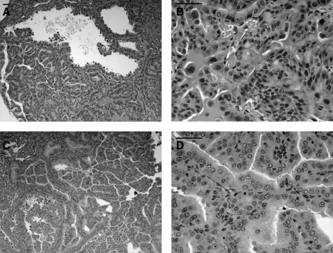
Figures

Similar articles
-
Lymphangioleiomyomatosis and TSC2-/- cells.Lymphat Res Biol. 2010 Mar;8(1):59-69. doi: 10.1089/lrb.2009.0031. Lymphat Res Biol. 2010. PMID: 20235888 Free PMC article. Review.
-
Rapamycin-insensitive up-regulation of adipocyte phospholipase A2 in tuberous sclerosis and lymphangioleiomyomatosis.PLoS One. 2014 Oct 27;9(10):e104809. doi: 10.1371/journal.pone.0104809. eCollection 2014. PLoS One. 2014. PMID: 25347447 Free PMC article.
-
Pathogenesis of multifocal micronodular pneumocyte hyperplasia and lymphangioleiomyomatosis in tuberous sclerosis and association with tuberous sclerosis genes TSC1 and TSC2.Pathol Int. 2001 Aug;51(8):585-94. doi: 10.1046/j.1440-1827.2001.01242.x. Pathol Int. 2001. PMID: 11564212 Review.
-
Mammalian target of rapamycin signaling and autophagy: roles in lymphangioleiomyomatosis therapy.Proc Am Thorac Soc. 2010 Feb;7(1):48-53. doi: 10.1513/pats.200909-104JS. Proc Am Thorac Soc. 2010. PMID: 20160148 Free PMC article. Review.
-
Urokinase-type plasminogen activator (uPA) is critical for progression of tuberous sclerosis complex 2 (TSC2)-deficient tumors.J Biol Chem. 2017 Dec 15;292(50):20528-20543. doi: 10.1074/jbc.M117.799593. Epub 2017 Sep 27. J Biol Chem. 2017. PMID: 28972182 Free PMC article.
Cited by
-
A vascular model of Tsc1 deficiency accelerates renal tumor formation with accompanying hemangiosarcomas.Mol Cancer Res. 2015 Mar;13(3):548-55. doi: 10.1158/1541-7786.MCR-14-0178. Epub 2014 Dec 29. Mol Cancer Res. 2015. PMID: 25548102 Free PMC article.
-
High Mobility Group AT-Hook 2 (HMGA2) Oncogenicity in Mesenchymal and Epithelial Neoplasia.Int J Mol Sci. 2020 Apr 29;21(9):3151. doi: 10.3390/ijms21093151. Int J Mol Sci. 2020. PMID: 32365712 Free PMC article. Review.
-
The neural crest lineage as a driver of disease heterogeneity in Tuberous Sclerosis Complex and Lymphangioleiomyomatosis.Front Cell Dev Biol. 2014 Nov 25;2:69. doi: 10.3389/fcell.2014.00069. eCollection 2014. Front Cell Dev Biol. 2014. PMID: 25505789 Free PMC article. Review.
-
Human Pluripotent Stem Cell-Derived TSC2-Haploinsufficient Smooth Muscle Cells Recapitulate Features of Lymphangioleiomyomatosis.Cancer Res. 2017 Oct 15;77(20):5491-5502. doi: 10.1158/0008-5472.CAN-17-0925. Epub 2017 Aug 22. Cancer Res. 2017. PMID: 28830860 Free PMC article.
-
Tuberous sclerosis complex 1: an epithelial tumor suppressor essential to prevent spontaneous prostate cancer in aged mice.Cancer Res. 2010 Nov 1;70(21):8937-47. doi: 10.1158/0008-5472.CAN-10-1646. Epub 2010 Oct 12. Cancer Res. 2010. PMID: 20940396 Free PMC article.
References
-
- Eker R. Familial renal adenomas in Wistar rats; A preliminary report. Acta Pathol Microbiol Scand. 1954;34:554–562. - PubMed
-
- Eker R. Mossige J. Johannessen JV. Aars H. Hereditary renal adenomas and adenocarcinomas in rats. Diagn Histopathol. 1981;4:99–110. - PubMed
-
- Everitt J. Goldsworthy T. Wolf D. Walker C. Hereditary renal cell carcinoma in the Eker rat: A rodent familial cancer syndrome. J Urol. 1992;148:1932–1936. - PubMed
Publication types
MeSH terms
Substances
Grants and funding
LinkOut - more resources
Full Text Sources
Other Literature Sources
Medical
Miscellaneous
