. 2011 Feb;17(1):4-31.
doi: 10.1111/j.1755-5949.2009.00116.x.

A review of the neural mechanisms of action and clinical efficiency of riluzole in treating amyotrophic lateral sclerosis: what have we learned in the last decade?

Affiliations

A review of the neural mechanisms of action and clinical efficiency of riluzole in treating amyotrophic lateral sclerosis: what have we learned in the last decade?

Mark C Bellingham. CNS Neurosci Ther. 2011 Feb.

Abstract

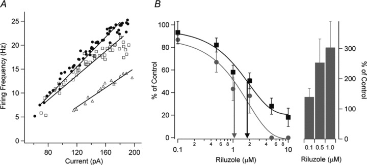
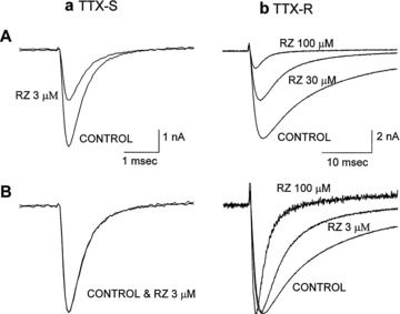
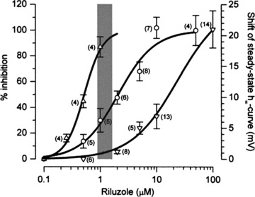
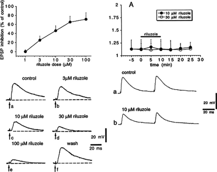
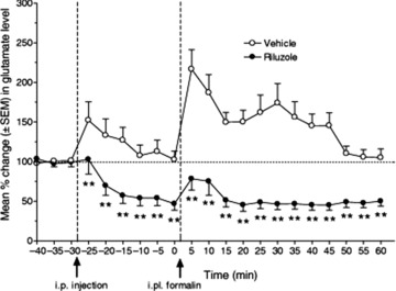
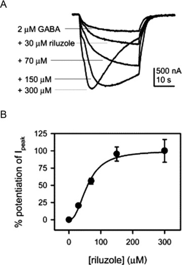
Amyotrophic lateral sclerosis (ALS) is a devastating and fatal neurodegenerative disease of adults which preferentially attacks the neuromotor system. Riluzole has been used as the only approved treatment for amyotrophic lateral sclerosis since 1995, but its mechanism(s) of action in slowing the progression of this disease remain obscure. Searching PubMed for "riluzole" found 705 articles published between January 1996 and June 2009. A systematic review of this literature found that riluzole had a wide range of effects on factors influencing neural activity in general, and the neuromotor system in particular. These effects occurred over a large dose range (<1 μM to >1 mM). Reported neural effects of riluzole included (in approximate ascending order of dose range): inhibition of persistent Na(+) current = inhibition of repetitive firing < potentiation of calcium-dependent K(+) current < inhibition of neurotransmitter release < inhibition of fast Na(+) current < inhibition of voltage-gated Ca(2+) current = promotion of neuronal survival or growth factors < inhibition of voltage-gated K(+) current = modulation of two-pore K(+) current = modulation of ligand-gated neurotransmitter receptors = potentiation of glutamate transporters. Only the first four of these effects commonly occurred at clinically relevant concentrations of riluzole (plasma levels of 1-2 μM with three- to four-fold higher concentrations in brain tissue). Treatment of human ALS patients or transgenic rodent models of ALS with riluzole most commonly produced a modest but significant extension of lifespan. Riluzole treatment was well tolerated in humans and animals. In animals, despite in vitro evidence that riluzole may inhibit rhythmic motor behaviors, in vivo administration of riluzole produced relatively minor effects on normal respiration parameters, but inhibited hypoxia-induced gasping. This effect may have implications for the management of hypoventilation and sleep-disordered breathing during end-stage ALS in humans.


Figures

Figure 1
Figure 1
Riluzole inhibits neuronal excitability and the persistent Na+ current (INaP) in cultured spinal neurons in a dose‐dependent way. (A) The relationship between firing frequency and current injection (F–I) and linear regressions are shown for control (•), 0.1 μM riluzole (□), and 1.0 μM riluzole (▵). The F–I gain is reduced and the current threshold for the onset of firing is increased with increasing riluzole concentrations. (B) The dose–response curve for all cells is shown for the effect of riluzole on the F–I gain (gray circle) for 0.1 μM riluzole (n = 11), 0.5 μM riluzole (n = 10), 1 μM riluzole (n = 8), 2 μM riluzole (n = 5), 5 μM riluzole (n = 5), and 10 μM riluzole (n = 5). The effect of riluzole on INaP (▪) is also shown for 0.1 μM riluzole (n = 6), 0.5 μM riluzole (n = 5), 1 μM riluzole (n = 7), 2 μM riluzole (n = 5), 5 μM riluzole (n = 5), and 10 μM riluzole (n = 5). The EC50 for riluzole inhibition of the F–I gain (gray arrow) was 1.1 μM and the EC50 for inhibition of INaP (black arrow) was 1.8 μM. Riluzole also dose‐dependently increased the current threshold for firing (right side, bars; mean ± SEM shown for [B]). Threshold amplitudes could not be measured above 2 μM riluzole because spiking behavior became very irregular. Reprinted from [17], copyright (2006), with permission from John Wiley & Sons.
Figure 2
Figure 2
Effects of riluzole (RZ) on tetrodotoxin‐sensitive (TTX‐S) and tetrodotoxin‐resistant (TTX‐R) sodium channel currents of rat dorsal root ganglion neurons. Currents were evoked by depolarizing steps to 0 mV from a holding potential of −80 mV. TTX‐S currents activated and inactivated rapidly and were selectively blocked by 200 nM TTX, whereas TTX‐R currents activated and inactivated more slowly and were unaffected by 200 nM TTX. (a) TTX‐sensitive sodium channel. (b) TTX‐resistant sodium channel. (A) Riluzole blocks TTX‐sensitive sodium channel currents more potently than TTX‐resistant sodium channel currents when the membrane was held at −80 mV. (B) Peak current amplitude in the presence of riluzole is normalized to the control current. Riluzole did not alter the activation and inactivation kinetics of TTX‐S currents, while the time course of inactivation of TTX‐R currents was accelerated. Reprinted from [44], copyright (1997), with permission from the American Society for Experimental Pharmacology and Therapeutics.
Figure 3
Figure 3
Overlaid view of the dose–response curves of riluzole on firing rate, INaP, and fast sodium current shows that decreases in firing are most likely due to inhibition of INaP. From left to right: concentration–response curves showing the effects of riluzole on firing rate (▵, left vertical axis), on INaP amplitude (○, left vertical axis), and on the shift of steady‐state inactivation curve for fast sodium current (∇, right vertical axis). The effect on firing rate was computed as the percentage ratio of the total number of events in 30 successive stimulations in tests over controls. For all concentrations INaP was recorded at the test potential of −15 mV (holding potential of −75 mV). Data relative to INaP were pooled after they had been normalized within each cell with respect to controls. For the shifts of the steady‐state inactivation curve (actually negative), absolute values are given. The gray band represents the putative plasma concentrations of riluzole [177] at the suggested therapeutic dose (2 × 50 mg/day). For all curves, data points are given as mean ± SEM, n within parentheses. Reprinted from [14], copyright (2000), with permission from John Wiley & Sons.
Figure 4
Figure 4
Riluzole inhibits non‐NMDA glutamate receptor excitatory postsynaptic potentials (EPSPs) in rat striatal spiny neurons but does not alter paired pulse facilitation. (Left panel) Riluzole inhibits EPSPs evoked by cortical stimulation. The graph in the upper part of the figure shows the dose–response curve obtained at various concentrations of riluzole on the amplitude of corticostriatal EPSPs. Each data point was obtained from at least four single experiments; the IC50 for EPSP inhibition was 6 μM. The lower part of the left panel shows averages (four single sweeps) of EPSPs recorded from a striatal spiny neuron under control condition, during the application of four different concentrations of riluzole and after 30 min washout. Each concentration was applied for 10 min. The resting membrane potential of the cell was −87 mV and was constant throughout the experiment. (Right panel) Riluzole does not alter paired pulse facilitation. (A) The graph shows the amplitude ratio of the second EPSP response to the first EPSP response (EPSP2:EPSP1) before, during, and after the application of two different concentrations of riluzole (black bar). Traces in the lower part of the figure show synaptic responses to paired stimulation under control condition (a) and after 10 min application of 10 μM riluzole (b). Reprinted from [12], copyright (1998), with permission from Elsevier.
Figure 6
Figure 6
Pretreatment with riluzole (12 mg/kg i.p.) produced significant antinociceptive effects following hind paw injection of formalin. The percentage change in glutamate content of in vivo microdialysate from rat thoracic spinal cord was measured by reverse phase HPLC with fluorescence detection. Riluzole (•) significantly reduced the formalin‐induced increase in spinal glutamate both in the first phase (from 5 to 15 min following i.p. injection of riluzole) and in the second phase (from 20 to 60 min) after formalin injection (**p < 0.01), compared to sham‐injected animals (○). Riluzole also significantly reduced basal spinal glutamate levels throughout the testing period. Reprinted from [129], copyright (2007), with permission of John Wiley & Sons.
Figure 5
Figure 5
Riluzole concentration‐dependently potentiated GABA responses from heterologously expressed receptors in Xenopus oocytes. (A) Sample recordings showing the potentiation of the response to 2 μM GABA induced by different concentrations of riluzole. With high concentrations of riluzole, GABA responses usually displayed apparent desensitization. (B) Concentration dependence of the effect of riluzole on the responses to 2 μM GABA. Normalized potentiation relative to the peak potentiation by 300 μM riluzole is plotted against riluzole concentration. Smooth curves are fit to the Hill equation. The Hill coefficient is 2.4, and riluzole EC50 (the concentration of riluzole that induces half‐maximal potentiation) is 58.7 μM (n = 5). Before normalization, the amplitudes of potentiation caused by different concentrations of riluzole were: 75.7 ± 7.1% from 30 μM riluzole, 208.0 ± 16.1% from 70 μM riluzole, 353.6 ± 38.1% from 150 μM riluzole, and 370.8 ± 61.5% from 300 μM riluzole. Reprinted from [104], copyright (2002), with permission from Elsevier.

Similar articles

Cited by

References

    1. Domino EF, Unna KR, Kerwin J. Pharmacological properties of benzazoles. I. Relationship between structure and paralyzing action. J Pharmacol Exp Ther 1952;105:486–497. - PubMed
    1. Doble A. The pharmacology and mechanism of action of riluzole. Neurology 1996;47:233S–241S. - PubMed
    1. Bruijn LI, Miller TM, Cleveland DW. Unraveling the mechanisms involved in motor neuron degeneration in ALS. Annu Rev Neurosci 2004;27:723–749. - PubMed
    1. Bensimon G, Ludolph A, Agid Y, et al Riluzole treatment, survival and diagnostic criteria in Parkinson plus disorders: The NNIPPS study. Brain 2009;132:156–171. - PMC - PubMed
    1. Landwehrmeyer GB, Dubois B, de Yebenes JG, et al Riluzole in Huntington's disease: A 3‐year, randomized controlled study. Ann Neurol 2007;62:262–272. - PubMed

Publication types