

Caudate atrophy on MRI is a characteristic feature of FTLD-FUS
- PMID: 20236174
- PMCID: PMC2989679
- DOI: 10.1111/j.1468-1331.2010.02975.x
Caudate atrophy on MRI is a characteristic feature of FTLD-FUS
Abstract
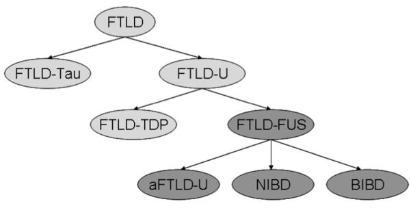
Background and purpose: Frontotemporal lobar degeneration (FTLD) can be subdivided into those in which the abnormal protein is tau (FTLD-TAU), the TAR DNA binding protein 43 (FTLD-TDP) and the fused in sarcoma protein (FTLD-FUS). We have observed severe caudate atrophy at autopsy in FTLD-FUS, and hence, we aimed to determine whether caudate atrophy on MRI is a feature that can distinguish FTLD-FUS from FTLD-TDP and FTLD-TAU.
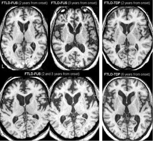
Methods: From a cohort of 207 cases of FTLD, we identified all cases of FTLD-FUS that had a volumetric antemortem head MRI (n = 3). Caudate and frontal lobe volumes were measured in all three cases using atlas-based parcellation and SPM5 and were compared to 10 randomly selected cases of FTLD-TDP and 10 randomly selected cases of FTLD-TAU. Total grey matter volumes were also calculated for all cases.
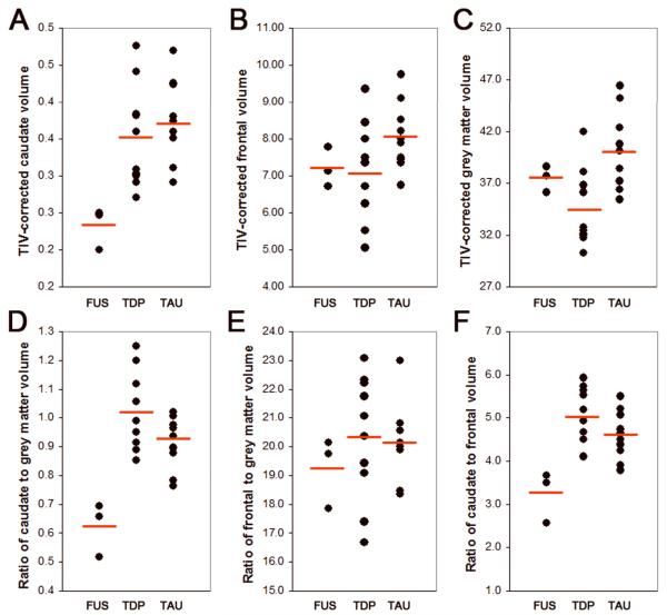
Results: The FTLD-FUS cases had significantly smaller caudate volumes (P = 0.02) yet similar frontal lobe grey matter volumes (P = 0.12) compared to FTLD-TDP and FTLD-TAU. Caudate volumes when corrected for total grey matter volume (P = 0.01) or frontal lobe grey matter volume (P = 0.01) were significantly smaller in FTLD-FUS than in FTLD-TDP and FTLD-TAU and showed no overlap with the other two groups.
Conclusions: Caudate atrophy on MRI appears to be significantly greater in FTLD-FUS compared with FTLD-TDP and FTLD-TAU, suggesting that severe caudate atrophy may be a useful clinical feature to predict FTLD-FUS pathology.
Figures
References
-
- Josephs KA. Frontotemporal dementia and related disorders: deciphering the enigma. Ann Neurol. 2008;64:4–14. - PubMed
-
- Neumann M, Sampathu DM, Kwong LK, et al. Ubiquitinated TDP-43 in frontotemporal lobar degeneration and amyotrophic lateral sclerosis. Science. 2006;314:130–133. - PubMed
-
- Josephs KA, Lin WL, Ahmed Z, Stroh DA, Graff-Radford NR, Dickson DW. Frontotemporal lobar degeneration with ubiquitin-positive, but TDP-43-negative inclusions. Acta Neuropathol. 2008;116:159–167. - PubMed
Publication types
MeSH terms
Substances
Grants and funding
LinkOut - more resources
Full Text Sources
