

Review
doi: 10.1111/j.1742-4658.2010.07609.x.
Epub 2010 Mar 4.
Mixed lineage leukemia: a structure-function perspective of the MLL1 protein
Affiliations
- PMID: 20236310
- PMCID: PMC2892832
- DOI: 10.1111/j.1742-4658.2010.07609.x
Item in Clipboard
Review
Mixed lineage leukemia: a structure-function perspective of the MLL1 protein
FEBS J.
2010 Apr.
Abstract
Several acute lymphoblastic and myelogenous leukemias are correlated with alterations in the human mixed lineage leukemia protein-1 (MLL1) gene. MLL1 is a member of the evolutionarily conserved SET1 family of histone H3 lysine 4 (H3K4) methyltransferases, which are required for the regulation of distinct groups of developmentally regulated genes in metazoans. Despite the important biological role of SET1 family enzymes and their involvement in human leukemias, relatively little is understood about how these enzymes work. Here we review several recent structural and biochemical studies that are beginning to shed light on the molecular mechanisms for the regulation of H3K4 methylation by the human MLL1 enzyme.
Figures

Schematic representation showing the domain architecture of the MLL1 protein. a) The full-length MLL1 protein is rapidly processed by the Taspase 1 enzyme into MLL-N and MLL-C fragments, which reassociate through FYRN and FYRC motifs to form a stable complex. This mature protein then assembles with a number of proteins to form MLL1 complexes in the cell. b) Known three-dimensional structures of conserved MLL1 domains (colored green in each image). On the top from left-to-right is the CXXC domain (PDB code: 2j2s), and the c-terminal SET domain (PDB code: 2w5z). On the bottom from left to right is the MLL1 TAD domain (green) bound to the CBP:c-Myb complex (orange and blue, respectively) (PDB code: 2agh); and the MLL1 Win motif (green) bound to the WDR5 protein (purple) (PDB code: 3eg6).

The CXXC and TAD domains of MLL1 help recruit MLL1 to target loci. a) Transparent surface representation of the MLL1 CXXC domain (purple) determined by heteronuclear NMR spectroscopy (PDB code: 2j2s). A cartoon of the protein backbone is shown with zinc ions represented as spheres. The surfaces of amino acid residues perturbed by DNA binding in chemical shift and mutagenesis experiments are indicated in blue. The location of the extended loop is indicated with an arrow. b) The CBP-KIX domain:cMyb binary complex. The CBP-KIX domain is shown in orange and the c-Myb transactivation domain is shown in blue (drawn from PDB code: 1sb0). The positions of E665 and E555 of the CBP-KIX domain, and residues K291 and K294 of the c-Myb tranactivation domain are indicated. c) The CBP-KIX:cMyb:MLL TAD domain ternary complex (drawn form PDB code: 2AGH). The MLL1 TAD domain is shown in green and the colors for the CBP-KIX:cMyb are as in a). Upon formation of the ternary complex, residues E665 and E666 of the CBP-KIX domain become ordered and interact with the c-Myb transactivation domain (indicated with the arrow).
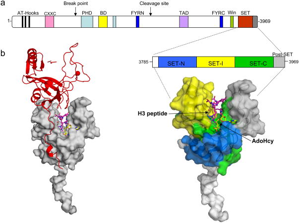
X-ray crystal structure of the c-terminal MLL1 SET domain bound to s-adenosyl homocysteine (yellow) and histone H3 peptide (purple) (PDB code: 2W5Z). (a) At the top is a schematic representation of the full-length MLL1 protein and blown up is the construct used for crystallization of the MLL1 SET domain (residues 3785–3969). The SET-N, SET-I, and SET-C sub-domains are colored in blue, yellow and green, respectively. The post-SET domain is colored in grey, and the N-flanking region is colored white. The positions of histone H3 and AdoHcy are indicated. b) Crystal packing constrains the MLL1 SET domain into an open conformation. Surface representation of the MLL1 SET domain (grey) shown with a symmetry related molecule in red. The N-terminus of the symmetry related molecule interacts extensively with the SET-I region-constraining the MLL1 SET domain in an open conformation.

X-ray crystal structure of the MLL1 Win motif peptide in complex with WDR5. At top the domain architecture of full-length MLL1 is shown. The blown up portion shows a cut-away view of the MLL1 Win motif (green) bound to the central opening of WDR5 (PDB code 3EG6). The position of the conserved Arg 3765 is indicated. On the left, a stick representation is used to show the position of the MLL1 Win motif residues 3762–3770 (green) bound to the central opening of WDR5. MLL Win motif residue numbers are indicated.

New model for the mechanism of multiple lysine methylation by the MLL1 core complex (adapted from [36]). a) The MLL1 core complex is composed of two distinct H3K4 methyltransferases each possessing H3K4 monomethylation activity on their own. The dashed oval on the WDR5-RbBP5-Ash2L-DPY-30 sub-complex indicates that the catalytic motif is presently unknown, and may be shared between subunits. b). WDR5’s recognition of the MLL1 Win motif results in the assembly of the MLL1 core complex, which possesses H3K4 dimethyltransferase activity. We suggest that the MLL1 SET domain catalyzes monomethylation of histone H3 at lysine 4, which is followed by transfer of the monomethylated histone H3 to a second active site on the WRAD sub-complex, where H3K4 dimethylation occurs. We propose that mechanisms that control the assembly of the MLL1 core complex will be important for the regulation of H3K4 methylation states in the cell.
References
-
- Leegte B, et al. 11q- syndrome: three cases and a review of the literature. Genet Couns. 1999;10(3):305–13. - PubMed
-
- Marschalek R. Mixed Lineage Leukemia: roles in human malignancies and potential therapy. FEBS J. 2010 - PubMed
-
- Yu BD, et al. Altered Hox expression and segmental identity in Mll-mutant mice. Nature. 1995;378(6556):505–8. - PubMed
Publication types
MeSH terms
Substances
Grants and funding
LinkOut - more resources
Full Text Sources
Research Materials
