

Reduced level of glutamic acid decarboxylase-67 kDa in the prefrontal cortex in major depression
- PMID: 20236554
- PMCID: PMC2857696
- DOI: 10.1017/S1461145709990587
Reduced level of glutamic acid decarboxylase-67 kDa in the prefrontal cortex in major depression
Abstract
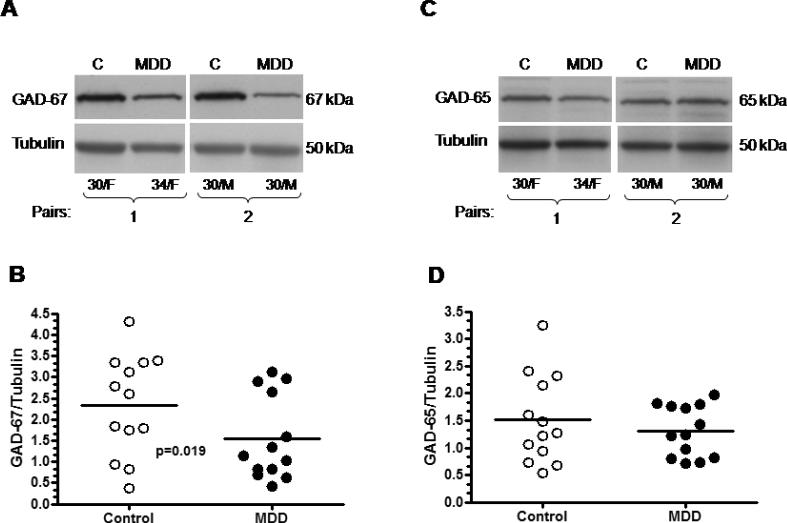
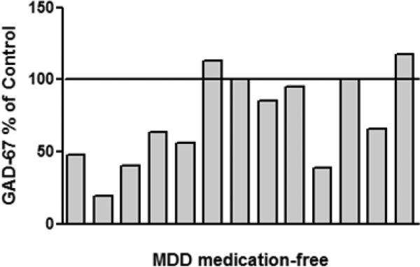
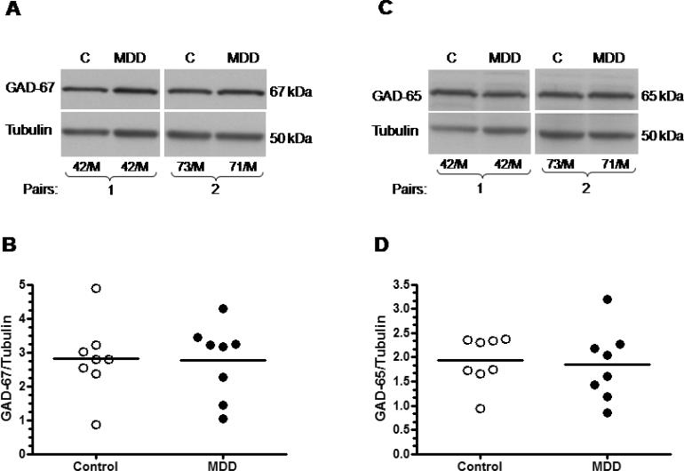
Accumulating evidence suggests dysfunction of the gamma-aminobutyric acid (GABA) system in major depressive disorder (MDD). Neuroimaging studies consistently report reductions of cortical GABA in depressed patients. Our post-mortem analyses demonstrate a reduction in the density and size of GABAergic interneurons in the dorsolateral prefrontal cortex (DLPFC) in MDD. The goal of this study was to test whether the level of glutamic acid decarboxylase (GAD), the GABA synthesizing enzyme, will also be reduced in the same cortical region in MDD. Levels of GAD-65 and GAD-67 proteins were investigated by Western blotting in samples from the DLPFC (BA 9) in 13 medication-free subjects with MDD, and 13 psychiatrically healthy controls. The overall amount of GAD-67 was significantly reduced (-34%) in depressed subjects compared to matched controls. Since recent neuroimaging studies have demonstrated that antidepressants modulate GABA levels, additional experiments were performed to examine the levels of GAD in eight depressed subjects treated with antidepressant medications. Levels of GAD-67 were unchanged in these depressed subjects compared to their respective controls (n=8). The overall amounts of GAD-65 were similar in depressed subjects compared to matched controls, regardless of antidepressant medication. Reduced levels of GAD-67, which is localized to somata of GABA neurons, further support our observation of a decreased density of GABAergic neurons in the PFC in depression. It is likely that a decrease in GAD-67 accounts for the reduction in GABA levels revealed by neuroimaging studies. Moreover, our data support previous neuroimaging observations that antidepressant medication normalizes GABA deficits in depression.
Figures
References
-
- American Psychiatric Association . Diagnostic and statistical manual of mental disorder. 4th edn. American Psychiatry Press; Washington DC: 1994.
-
- Asada H, Kawamura Y, Maruyama K, Kume H, Ding R, Ji FY, Kanbara N, Kuzume H, Sanbo M, Yagi T, Obata K. Mice lacking the 65 kDa isoform of glutamic acid decarboxylase (GAD65) maintain normal levels of GAD67 and GABA in their brains but are susceptible to seizures. Biochemical and Biophysical Research Communications. 1996;229:891–895. - PubMed
-
- Asada H, Kawamura Y, Maruyama K, Kume H, Ding RG, Kanbara N, Kuzume H, Sanbo M, Yagi T, Obata K. Cleft palate and decreased brain gamma-aminobutyric acid in mice lacking the 67-kDa isoform of glutamic acid decarboxylase. Proceedings of the National Academy of Sciences of the United States of America. 1997;94:6496–6499. - PMC - PubMed
-
- Bhagwagar Z, Wylezinska M, Taylor M, Jezzard P, Matthews PM, Cowen PJ. Increased brain GABA concentrations following acute administration of a selective serotonin reuptake inhibitor. American Journal of Psychiatry. 2004;161:368–370. - PubMed
-
- Bowdler JM, Green AR, Minchin MC, Nutt DJ. Regional GABA concentration and [3H]-diazepam binding in rat brain following repeated electroconvulsive shock. Journal of Neural Transmission. 1983;56:3–12. - PubMed
Publication types
MeSH terms
Substances
Grants and funding
LinkOut - more resources
Full Text Sources
Miscellaneous
