

'Let the phage do the work': using the phage P22 coat protein structures as a framework to understand its folding and assembly mutants
- PMID: 20236676
- PMCID: PMC2862144
- DOI: 10.1016/j.virol.2010.02.017
'Let the phage do the work': using the phage P22 coat protein structures as a framework to understand its folding and assembly mutants
Abstract
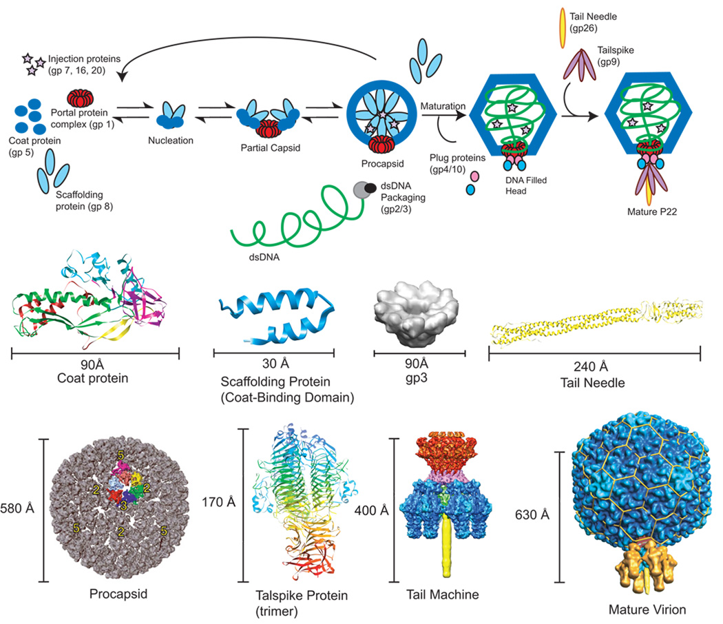
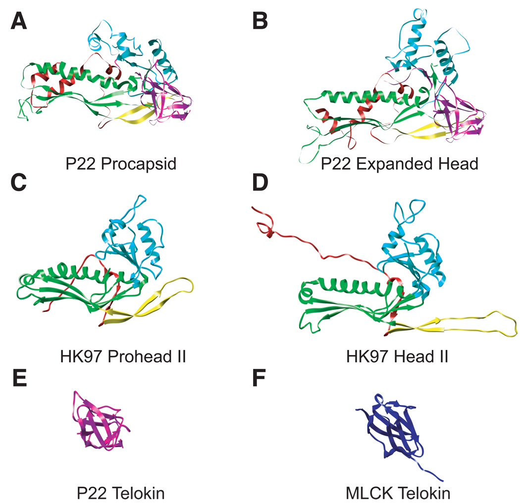
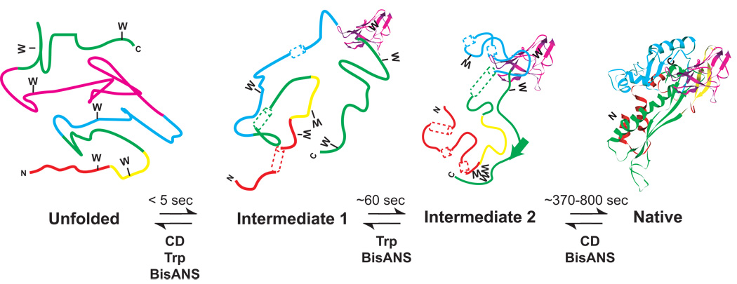
The amino acid sequence of viral capsid proteins contains information about their folding, structure and self-assembly processes. While some viruses assemble from small preformed oligomers of coat proteins, other viruses such as phage P22 and herpesvirus assemble from monomeric proteins (Fuller and King, 1980; Newcomb et al., 1999). The subunit assembly process is strictly controlled through protein:protein interactions such that icosahedral structures are formed with specific symmetries, rather than aberrant structures. dsDNA viruses commonly assemble by first forming a precursor capsid that serves as a DNA packaging machine (Earnshaw, Hendrix, and King, 1980; Heymann et al., 2003). DNA packaging is accompanied by a conformational transition of the small precursor procapsid into a larger capsid for isometric viruses. Here we highlight the pseudo-atomic structures of phage P22 coat protein and rationalize several decades of data about P22 coat protein folding, assembly and maturation generated from a combination of genetics and biochemistry.
2010 Elsevier Inc. All rights reserved.
Figures
Similar articles
-
A Molecular Staple: D-Loops in the I Domain of Bacteriophage P22 Coat Protein Make Important Intercapsomer Contacts Required for Procapsid Assembly.J Virol. 2015 Oct;89(20):10569-79. doi: 10.1128/JVI.01629-15. Epub 2015 Aug 12. J Virol. 2015. PMID: 26269173 Free PMC article.
-
Highly specific salt bridges govern bacteriophage P22 icosahedral capsid assembly: identification of the site in coat protein responsible for interaction with scaffolding protein.J Virol. 2014 May;88(10):5287-97. doi: 10.1128/JVI.00036-14. Epub 2014 Mar 5. J Virol. 2014. PMID: 24600011 Free PMC article.
-
The role of the coat protein A-domain in p22 bacteriophage maturation.Viruses. 2014 Jul 14;6(7):2708-22. doi: 10.3390/v6072708. Viruses. 2014. PMID: 25025835 Free PMC article.
-
Nature's favorite building block: Deciphering folding and capsid assembly of proteins with the HK97-fold.Virology. 2015 May;479-480:487-97. doi: 10.1016/j.virol.2015.02.055. Epub 2015 Apr 8. Virology. 2015. PMID: 25864106 Free PMC article. Review.
-
Virus particle maturation: insights into elegantly programmed nanomachines.Curr Opin Struct Biol. 2010 Apr;20(2):210-6. doi: 10.1016/j.sbi.2010.01.004. Epub 2010 Feb 9. Curr Opin Struct Biol. 2010. PMID: 20149636 Free PMC article. Review.
Cited by
-
Intravirion DNA Can Access the Space Occupied by the Bacteriophage P22 Ejection Proteins.Viruses. 2021 Jul 30;13(8):1504. doi: 10.3390/v13081504. Viruses. 2021. PMID: 34452369 Free PMC article.
-
Architecture of the Complex Formed by Large and Small Terminase Subunits from Bacteriophage P22.J Mol Biol. 2015 Oct 9;427(20):3285-3299. doi: 10.1016/j.jmb.2015.08.013. Epub 2015 Aug 21. J Mol Biol. 2015. PMID: 26301600 Free PMC article.
-
Multiple functional roles of the accessory I-domain of bacteriophage P22 coat protein revealed by NMR structure and CryoEM modeling.Structure. 2014 Jun 10;22(6):830-41. doi: 10.1016/j.str.2014.04.003. Epub 2014 May 15. Structure. 2014. PMID: 24836025 Free PMC article.
-
Design of a VLP-nanovehicle for CYP450 enzymatic activity delivery.J Nanobiotechnology. 2015 Oct 9;13:66. doi: 10.1186/s12951-015-0127-z. J Nanobiotechnology. 2015. PMID: 26452461 Free PMC article.
-
The energetic contributions of scaffolding and coat proteins to the assembly of bacteriophage procapsids.Virology. 2012 Jun 20;428(1):64-9. doi: 10.1016/j.virol.2012.03.017. Epub 2012 Apr 20. Virology. 2012. PMID: 22520942 Free PMC article.
References
-
- Anderson E, Teschke CM. Folding of Phage P22 Coat Protein Monomers: Kinetic and Thermodynamic Properties. Virology. 2003;313:184–197. - PubMed
-
- Aramli L, Teschke C. Alleviation of a defect in protein folding by increasing the rate of subunit assembly. J Biol Chem. 2001;276(27):25372–25377. - PubMed
-
- Aramli LA, Teschke CM. Single amino acid substitutions globally suppress the folding defects of temperature-sensitive folding mutants of phage P22 coat protein. J. Biol. Chem. 1999;274(32):22217–22224. - PubMed
-
- Bamford DH, Grimes JM, Stuart DI. What does structure tell us about virus evolution? Curr Opin Struct Biol. 2005;15:655–63. - PubMed
-
- Bazinet C, King J. Initiation of P22 procapsid assembly in vivo. J. Mol. Biol. 1988;202(1):77–86. - PubMed
Publication types
MeSH terms
Substances
Grants and funding
LinkOut - more resources
Full Text Sources
Other Literature Sources
