. 2010 Dec;29(6):824-31.
doi: 10.1016/j.clnu.2010.02.009. Epub 2010 Mar 16.

Abscisic acid ameliorates experimental IBD by downregulating cellular adhesion molecule expression and suppressing immune cell infiltration

Affiliations

Abscisic acid ameliorates experimental IBD by downregulating cellular adhesion molecule expression and suppressing immune cell infiltration

Amir J Guri et al. Clin Nutr. 2010 Dec.

Abstract

Background & aims: Abscisic acid (ABA) has shown effectiveness in ameliorating inflammation in obesity, diabetes and cardiovascular disease models. The objective of this study was to determine whether ABA prevents or ameliorates experimental inflammatory bowel disease (IBD).

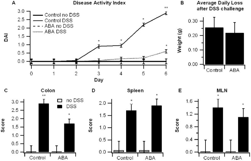
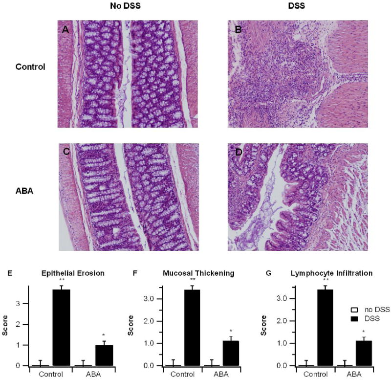
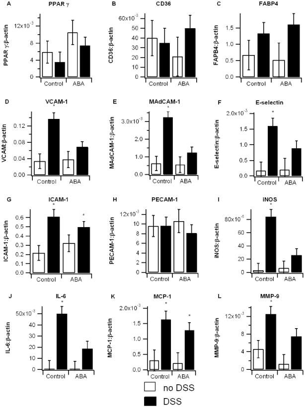
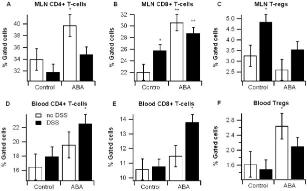
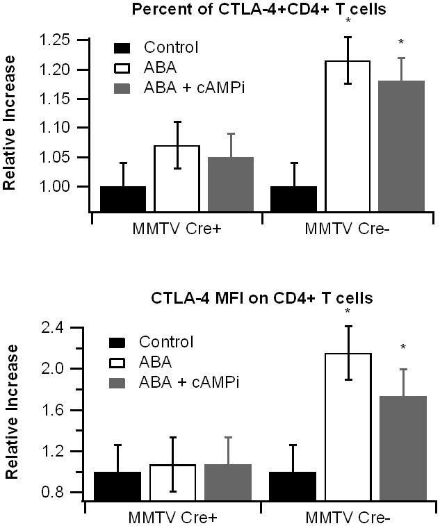
Methods: C57BL/6J mice were fed diets with or without ABA (100mg/kg) for 35 days prior to challenge with 2.5% dextran sodium sulfate (DSS). The severity of clinical disease was assessed daily. Colonic mucosal lesions were evaluated by histopathology, and cellular adhesion molecular and inflammatory markers were assayed by real-time quantitative PCR. Flow cytometry was used to quantify leukocyte populations in the blood, spleen, and mesenteric lymph nodes (MLN). The effect of ABA on cytotoxic T-lymphocyte antigen 4 (CTLA-4) expression in splenocytes was also investigated.

Results: ABA significantly ameliorated disease activity, colitis and reduced colonic leukocyte infiltration and inflammation. These improvements were associated with downregulation in vascular cell adhesion marker-1 (VCAM-1), E-selectin, and mucosal addressin adhesion marker-1 (MAdCAM-1) expression. ABA also increased CD4(+) and CD8(+) T-lymphocytes in blood and MLN and regulatory T cells in blood. In vitro, ABA increased CTLA-4 expression through a PPAR γ-dependent mechanism.

Conclusions: We conclude that ABA ameliorates gut inflammation by modulating T cell distribution and adhesion molecule expression.


Conflict of interest statement

Conflict of interest statement No conflicts to report

Figures

Figure 1
Figure 1
Effect of dietary abscisic acid (ABA) supplementation on disease severity. C57BL/6J mice were fed ABA-supplemented (100 mg/kg) or control diets for 35 days and challenged with 2.5% dextran sodium sulfate (DSS) or water (no DSS) for 7 days. The disease activity index (DAI), a composite score reflecting clinical signs of the disease (i.e. perianal soiling, rectal bleeding, diarrhea, and piloerection) was assessed daily (A) and the average daily loss in body weights (B) were calculated for mice undergoing the DSS challenge. On day 7 mice were euthanized and the colon, spleen, and mesenteric lymph nodes (MLN) (C-E) were macroscopically scored for inflammation. Data are represented as mean ± standard error. Points with an asterisk are significantly different from control, non-DSS treatment (P<0.05).
Figure 2
Figure 2
Effect of dietary abscisic acid (ABA) supplementation on colon histopathology. C57BL/6J mice were fed ABA-supplemented (100 mg/kg) or control diets for 35 days and challenged with 2.5% dextran sodium sulfate (DSS) or water (no DSS) for 7 days. Representative photomicrographs from control, no-DSS (A), control, DSS (B), ABA, no-DSS (C), and ABA, DSS (D) groups are depicted. All specimens underwent blinded histological examination and were scored (1-4) on epithelial erosion (E), mucosal wall thickening (F), and leukocyte infiltration (G). Data are represented as mean ± standard error. Points with an asterisk are significantly different from control, non-DSS treatment (P<0.05).
Figure 3
Figure 3
Effect of dietary abscisic acid (ABA) supplementation on colon gene expression. C57BL/6J mice were fed ABA-supplemented (100 mg/kg) or control diets for 35 days and challenged with 2.5% dextran sodium sulfate (DSS) or water (no DSS) for 7 days. Expressions of peroxisome proliferator-activated receptor γ (PPAR γ) and PPAR γ-responsive genes CD36, fatty acid binding protein 4 (FABP-4) (A-C), adhesion molecules vascular adhesion molecule 1 (VCAM-1), muscosal vascular addressin cell adhesion molecule 1 (MAdCAM-1), E-selectin, intracellular adhesion molecule 1 (ICAM-1), and platelet/endothelial cell adhesion molecule 1 (PECAM-1) (D-H), and pro-inflammatory proteins interleukin-6 (IL-6), inducible nitric oxide synthase (iNOS), monocyte chemoattractant protein-1 (MCP-1), and matrix metalloproteinase 9 (MMP-9) (I-L) were assessed by real-time quantitative PCR. Data are represented as mean ± standard error. Points with an asterisk are significantly different from control, non-DSS treatment (P<0.05).
Figure 4
Figure 4
Effect of dietary abscisic acid (ABA) supplementation on lymphocyte subsets in mesenteric lymph nodes (MLN) and blood. MLN (A-C) and blood (D-E) were immunophenotyped with anti-CD3, anti-CD4, anti-CD8, and anti-FoxP3 mouse monoclonal antibodies and the lymphocyte populations were analyzed with flow cytometry with FACS diva software. Data are represented as mean ± standard error. Points with an asterisk are significantly different from control, non-DSS treatment (P<0.05).
Figure 5
Figure 5
Effect of abscisic acid (ABA) treatment on cytotoxic T-lymphocyte antigen 4(CTLA-4) expression. Splenocytes were isolated from peroxisome proliferator-activated receptor γ (PPAR γ)-flfl; MMTV-Cre+ mice, who lack PPAR γ in hematopoietic cells, and control MMTV-Cre- littermates. Cells were stimulated with anti-CD3 (5 μg/mL)/CD28 (1 μg/mL) and treated with ABA (10 μM with or without the cAMP inhibitor 2’5’-dideoxyadenosine (icAMP, 10 μM) or 14-22 myristolated PKA inhibitor fragment (iPKA, 30 μM), a PKA-specific inhibitor, for 20 hours. Expression of CTLA-4 on CD3+, CD4+, and CD8+ lymphocytes was assessed with flow cytometry. Representative histograms (A-D) of CTLA-4 expression on CD3+ cells in MMTV, Cre- cells are depicted. The percentage of CTLA-4+ cells and CTLA-4 median fluorescence intensity (MFI) in CD8+ (E-F) and CD4+ (G-H) in PPAR γ expressing (black bars) and non-expressing cells (white bars) were assessed. Data are represented as mean ± standard error. Points with an asterisk indicate a significant effect of the treatment in comparison to the DMSO control (P<0.05).

Similar articles

Cited by

References

    1. Hanauer SB. Inflammatory bowel disease: epidemiology, pathogenesis, and therapeutic opportunities. Inflamm Bowel Dis. 2006;12(Suppl 1):S3–9. - PubMed
    1. Hanauer SB, Present DH. The state of the art in the management of inflammatory bowel disease. Rev Gastroenterol Disord. 2003;3(2):81–92. - PubMed
    1. Mackner LM, Sisson DP, Crandall WV. Review: psychosocial issues in pediatric inflammatory bowel disease. J Pediatr Psychol. 2004;29(4):243–257. - PubMed
    1. Bassaganya-Riera J, Skoneczka J, Kingston DGJ, Krishnan A, Misyak SA, Carter A, Pereira A, Guri AJ, Minorsky P, Turmakin R, et al. Mechanisms of action and medicinal applications of abscisic acid. Current Medicinal Chemistry. 2009 In press. - PubMed
    1. Fan J, Hill L, Crooks C, Doerner P, Lamb C. Abscisic acid has a key role in modulating diverse plant-pathogen interactions. Plant Physiol. 2009;150(4):1750–1761. - PMC - PubMed

Publication types

MeSH terms