

Chronic myofascial temporomandibular pain is associated with neural abnormalities in the trigeminal and limbic systems
- PMID: 20236763
- PMCID: PMC2860657
- DOI: 10.1016/j.pain.2010.01.006
Chronic myofascial temporomandibular pain is associated with neural abnormalities in the trigeminal and limbic systems
Abstract
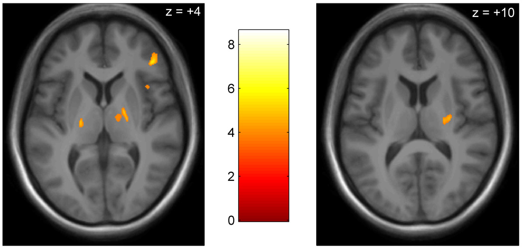
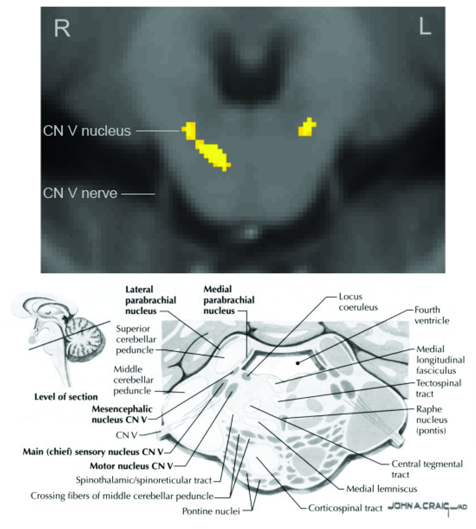
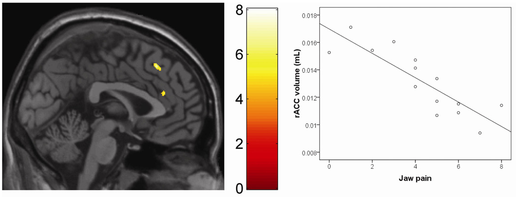
Myofascial pain of the temporomandibular region (M-TMD) is a common, but poorly understood chronic disorder. It is unknown whether the condition is a peripheral problem, or a disorder of the central nervous system (CNS). To investigate possible CNS substrates of M-TMD, we compared the brain morphology of 15 women with M-TMD to that of 15 age- and gender-matched healthy controls. High-resolution structural brain and brainstem scans were carried out using magnetic resonance imaging (MRI), and data were analyzed using a voxel-based morphometry approach. The M-TMD group evidenced decreased or increased gray matter volume compared to controls in several areas of the trigeminothalamocortical pathway, including brainstem trigeminal sensory nuclei, the thalamus, and the primary somatosensory cortex. In addition, M-TMD individuals showed increased gray matter volume compared to controls in limbic regions such as the posterior putamen, globus pallidus, and anterior insula. Within the M-TMD group, jaw pain, pain tolerance, and pain duration were differentially associated with brain and brainstem gray matter volume. Self-reported pain severity was associated with increased gray matter in the rostral anterior cingulate cortex and posterior cingulate. Sensitivity to pressure algometry was associated with decreased gray matter in the pons, corresponding to the trigeminal sensory nuclei. Longer pain duration was associated with greater gray matter in the posterior cingulate, hippocampus, midbrain, and cerebellum. The pattern of gray matter abnormality found in M-TMD individuals suggests the involvement of trigeminal and limbic system dysregulation, as well as potential somatotopic reorganization in the putamen, thalamus, and somatosensory cortex.
Copyright 2010 International Association for the Study of Pain. Published by Elsevier B.V. All rights reserved.
Conflict of interest statement
The authors have no conflicts of interest to disclose.
Figures
Similar articles
-
The brain in chronic CRPS pain: abnormal gray-white matter interactions in emotional and autonomic regions.Neuron. 2008 Nov 26;60(4):570-81. doi: 10.1016/j.neuron.2008.08.022. Neuron. 2008. PMID: 19038215 Free PMC article.
-
Changes in regional gray and white matter volume in patients with myofascial-type temporomandibular disorders: a voxel-based morphometry study.J Orofac Pain. 2011 Spring;25(2):99-106. J Orofac Pain. 2011. PMID: 21528116
-
Different pain, different brain: thalamic anatomy in neuropathic and non-neuropathic chronic pain syndromes.J Neurosci. 2011 Apr 20;31(16):5956-64. doi: 10.1523/JNEUROSCI.5980-10.2011. J Neurosci. 2011. PMID: 21508220 Free PMC article.
-
Age-related changes in the structure and function of brain regions involved in pain processing.Pain Med. 2012 Apr;13 Suppl 2:S37-43. doi: 10.1111/j.1526-4637.2011.01287.x. Pain Med. 2012. PMID: 22497746 Review.
-
Anatomy of the brainstem: a gaze into the stem of life.Semin Ultrasound CT MR. 2010 Jun;31(3):196-219. doi: 10.1053/j.sult.2010.03.006. Semin Ultrasound CT MR. 2010. PMID: 20483389 Review.
Cited by
-
Topology of pain networks in patients with temporomandibular disorder and pain-free controls with and without concurrent experimental pain: A pilot study.Front Pain Res (Lausanne). 2022 Oct 17;3:966398. doi: 10.3389/fpain.2022.966398. eCollection 2022. Front Pain Res (Lausanne). 2022. PMID: 36324873 Free PMC article.
-
Mindfulness-Based Stress Reduction, Cognitive Behavioral Therapy, and Acupuncture in Chronic Low Back Pain: Protocol for Two Linked Randomized Controlled Trials.JMIR Res Protoc. 2022 Sep 27;11(9):e37823. doi: 10.2196/37823. JMIR Res Protoc. 2022. PMID: 36166279 Free PMC article.
-
HIV Distal Neuropathic Pain Is Associated with Smaller Ventral Posterior Cingulate Cortex.Pain Med. 2017 Mar 1;18(3):428-440. doi: 10.1093/pm/pnw180. Pain Med. 2017. PMID: 27497320 Free PMC article.
-
Pain sensitivity is inversely related to regional grey matter density in the brain.Pain. 2014 Mar;155(3):566-573. doi: 10.1016/j.pain.2013.12.004. Epub 2013 Dec 11. Pain. 2014. PMID: 24333778 Free PMC article.
-
Rapid treatment-induced brain changes in pediatric CRPS.Brain Struct Funct. 2016 Mar;221(2):1095-111. doi: 10.1007/s00429-014-0957-8. Epub 2014 Dec 17. Brain Struct Funct. 2016. PMID: 25515312 Free PMC article.
References
-
- Alanen P. Occlusion and temporomandibular disorders (TMD): still unsolved question? J Dent Res. 2002;81:518–519. - PubMed
-
- Ashburner J. A fast diffeomorphic image registration algorithms. Neuroimage. 2007;38:95–113. - PubMed
-
- Bingel U, Gläscher J, Weiller C, Büchel C. Somatotopic representation of nociceptive information in the putamen: an event-related fMRI study. Cereb Cortex. 2004;14:1340–1345. - PubMed
Publication types
MeSH terms
Grants and funding
LinkOut - more resources
Full Text Sources
