. 2010 Jul;150(1):29-40.
doi: 10.1016/j.pain.2010.02.016. Epub 2010 Mar 16.

Selectively targeting pain in the trigeminal system

Affiliations

Selectively targeting pain in the trigeminal system

Hyun Yeong Kim et al. Pain. 2010 Jul.

Abstract

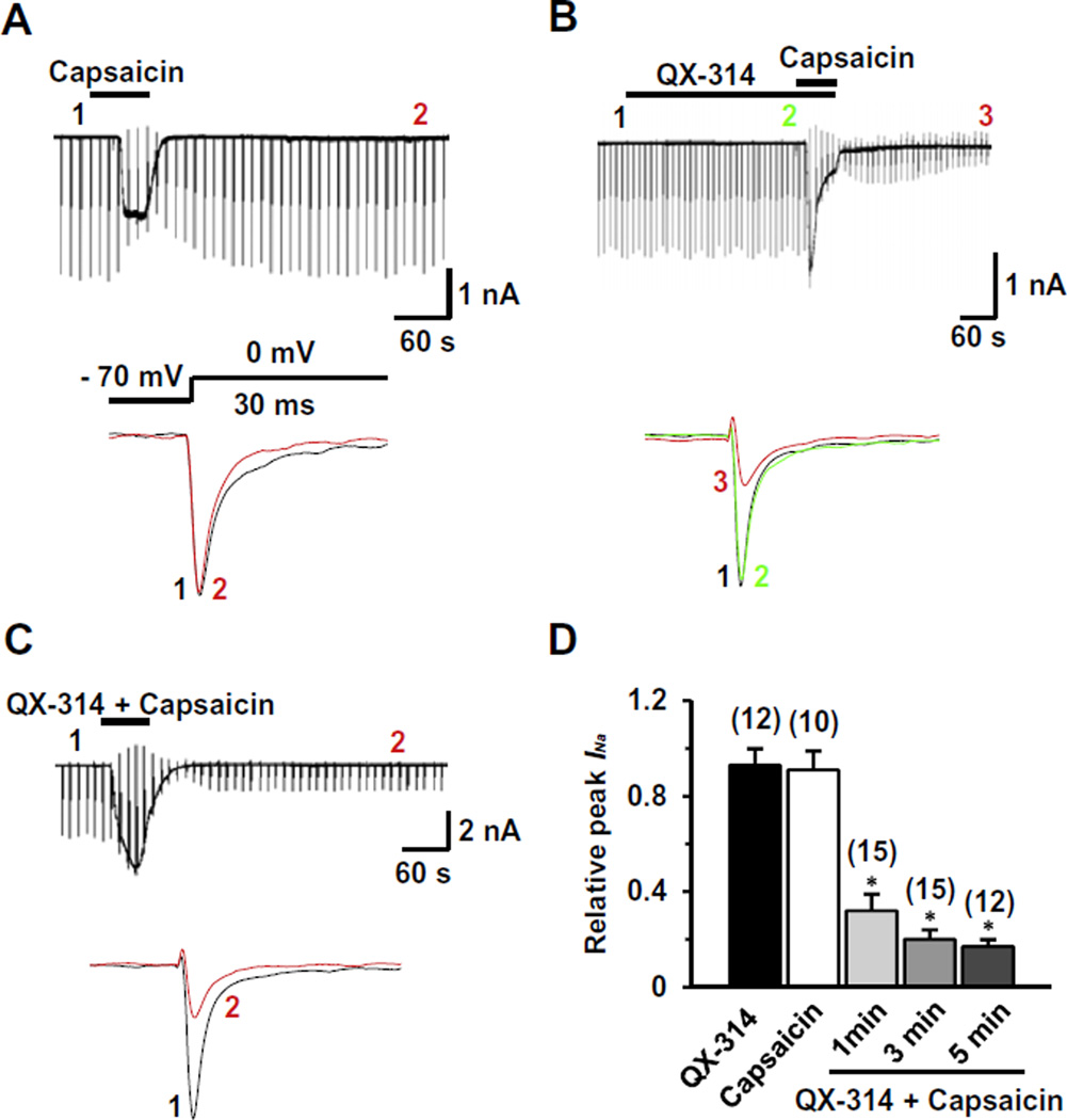
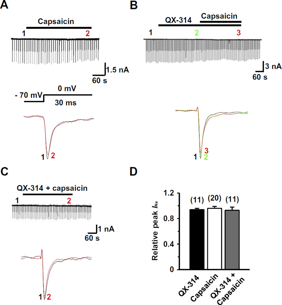
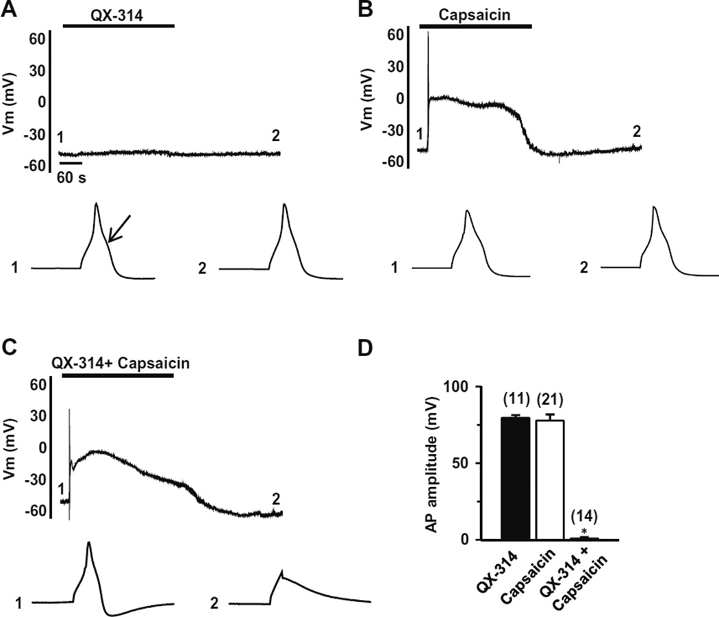
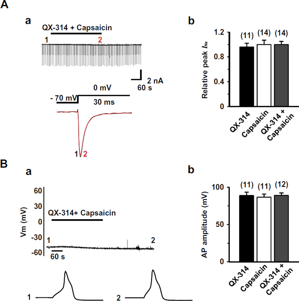
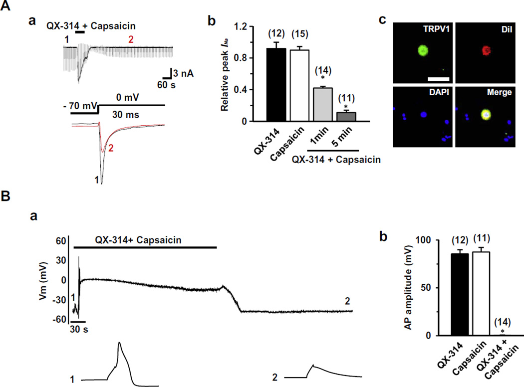
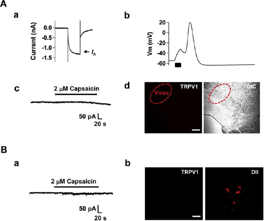
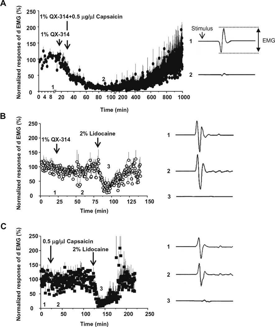
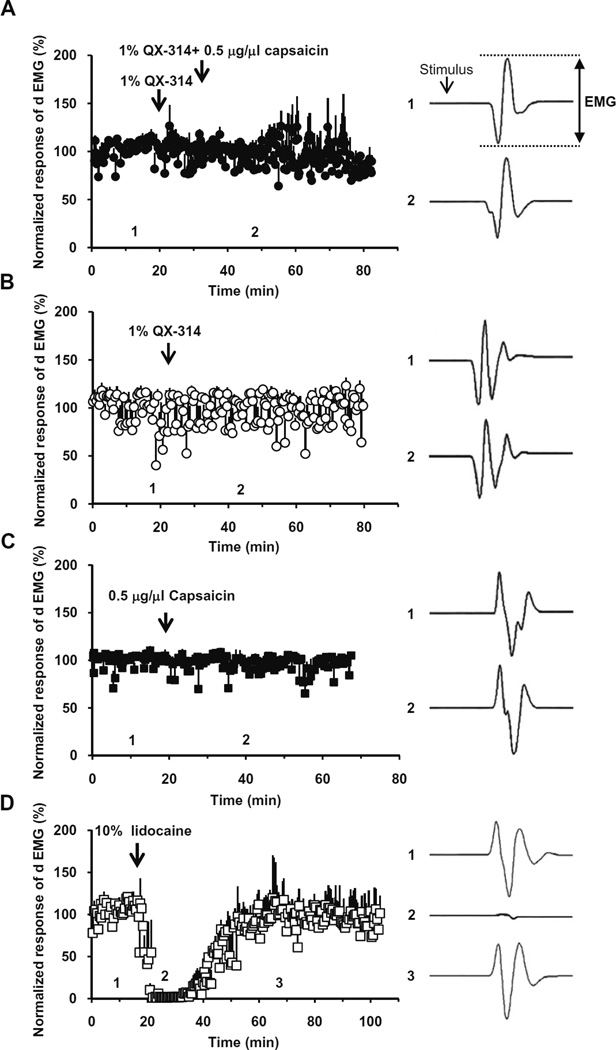
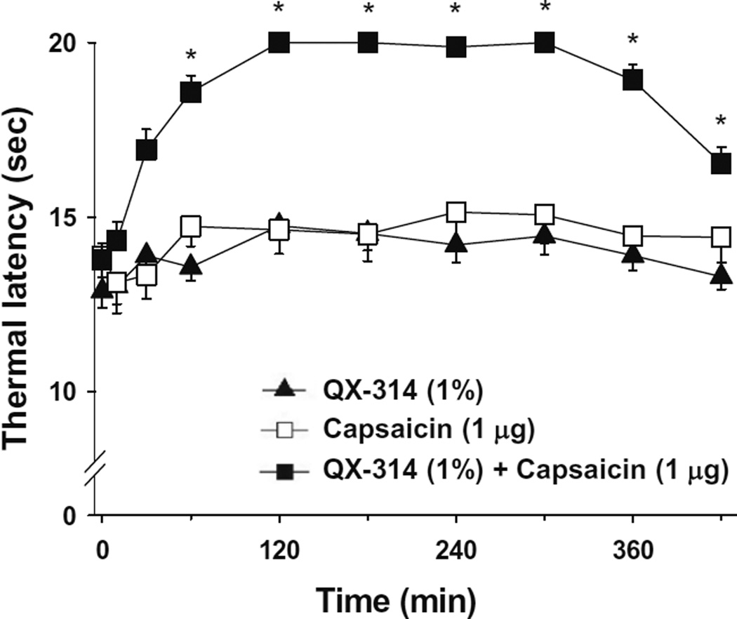
We tested whether it is possible to selectively block pain signals in the orofacial area by delivering the permanently charged lidocaine derivative QX-314 into nociceptors via TPRV1 channels. We examined the effects of co-applied QX-314 and capsaicin on nociceptive, proprioceptive, and motor function in the rat trigeminal system. QX-314 alone failed to block voltage-gated sodium channel currents (I(Na)) and action potentials (APs) in trigeminal ganglion (TG) neurons. However, co-application of QX-314 and capsaicin blocked I(Na) and APs in TRPV1-positive TG and dental nociceptive neurons, but not in TRPV1-negative TG neurons or in small neurons from TRPV1 knock-out mice. Immunohistochemistry revealed that TRPV1 is not expressed by trigeminal motor and trigeminal mesencephalic neurons. Capsaicin had no effect on rat trigeminal motor and proprioceptive mesencephalic neurons and therefore should not allow QX-314 to enter these cells. Co-application of QX-314 and capsaicin inhibited the jaw-opening reflex evoked by noxious electrical stimulation of the tooth pulp when applied to a sensory but not a motor nerve, and produced long-lasting analgesia in the orofacial area. These data show that selective block of pain signals can be achieved by co-application of QX-314 with TRPV1 agonists. This approach has potential utility in the trigeminal system for treating dental and facial pain.


Figures

Fig. 1
Fig. 1
Effects of QX-314 and capsaicin on voltage-gated sodium currents (INa) in capsaicin-sensitive TG neurons from adult rats. Representative recordings following the extracellular application of capsaicin alone (1 µM, 1 min) (A), co-application of QX-314 (5 mM) and capsaicin (1 min) after pretreatment by QX-314 alone (B) and co-application of QX-314 and capsaicin (1 min) (C). Upper panel: Long time-base recordings of INa measured during 30-ms voltage steps from a holding potential of −70 mV to a test potential of 0 mV delivered every 10 s. Each vertical deflection indicates INa measured every 10 s. Capsaicin, QX-314 or QX-314 plus capsaicin was applied during the time indicated by the horizontal bar. Lower panel: fast time-base recordings of superimposed sodium current evoked by test pulse at the points indicated in the upper panel. (D) Collected results for the effects of QX-314, capsaicin, or co-application of both on INa in capsaicin-sensitive TG neurons. The numbers in parentheses indicate the number of cells tested. Results are means ± S.E.M. *P < 0.05 compared with the control (INa peak before drug application).
Fig. 2
Fig. 2
Effects of QX-314 and capsaicin on INa in capsaicin-insensitive rat TG neurons. Representative recordings following the extracellular application of capsaicin alone (1 µM) (A), co-application of QX-314 (5 mM) and capsaicin with the pretreatment of QX-314 (B) and co-application of QX-314 and capsaicin (C). Upper panel: Long time-base recordings of INa measured during 30-ms voltage steps from a holding potential of −70 mV to a test potential of 0 mV delivered every 10 s. Each vertical deflection indicates INa measured every 10 s. Capsaicin, QX-314 or QX-314 plus capsaicin was applied during the time indicated by the horizontal bar. Lower panel: fast time-base recordings of superimposed sodium current evoked by test pulse at the points indicated in the upper panel. (D) Collected results for the effects of QX-314, capsaicin, or co-application of both on INa in capsaicin-sensitive TG neurons. The numbers in parentheses indicate the number of cells tested. Results are means ± S.E.M. *P < 0.05 compared with the control (INa peak before drug application).
Fig. 3
Fig. 3
Effects of QX-314 and capsaicin on action potentials in capsaicin-sensitive TG neurons. Representative current-clamp recordings over time following the application of QX-314 (5 mM, 5 min) (A), capsaicin (1 µM, 5 min) (B) or co-application of capsaicin and QX-314 (5 min) (C). Upper panel: action potentials (APs) were elicited by injection of 3-ms depolarizing current pulses with 250 pA amplitude. Note that membrane depolarization and transient action potential discharges were evoked by capsaicin in the capsaicin-sensitive TG neurons. Lower panel: APs recorded at the time points indicated at each upper panel. Arrow indicates the typical hump in the falling phase of AP in the capsaicin-sensitive TG neurons. (D) Collected results for the effects of QX-314, capsaicin, or co-application of both on AP amplitude in capsaicin-sensitive TG neurons. The numbers in parentheses indicate the number of cells tested. Results are means ± S.E.M. *P < 0.05 compared with the control (AP amplitude before drug application).
Fig. 4
Fig. 4
Effect of co-application of capsaicin and QX-314 on INa and APs in TRPV1 knock-out mice. (A) (a) Upper panel: a representative recording following the application or co-application of QX-314 (5 mM) and capsaicin (1 µM, 5 min) in the TRPV1 knock-out mice TG neurons. Lower panel: superimposed INa evoked by test pulse at the points indicated in upper panel. (b) Summary of the effects of QX-314, capsaicin, or co-application of both on INa in TG neurons from TRPV1 knock-out mice. (B) (a) Upper panel: A representative recording following the application or co-application of QX-314 (5 mM) and capsaicin (1 µM, 5 min). Lower panel: APs recorded at the time points indicated at the upper panel. (b) Collected results for the effects of QX-314, capsaicin, or co-application of both on AP amplitude in TRPV1 knock-out mice. TG neurons were small sized (<25 µm in diameter) with a resting membrane potential (Vres) of −54.2 ± 4.6 mV. The numbers in parentheses indicate the number of cells tested. Results are means ± S.E.M.
Fig. 5
Fig. 5
Effect of co-application of capsaicin and QX-314 on INa and APs in nociceptive dental primary afferent neurons. (A) (a) Upper panel: a representative recordings following the application or co-application of QX-314 (5 mM) and capsaicin (1 µM, 1 min) in dental primary afferent neurons. Lower panel: superimposed INa evoked by test pulse at the points indicated at the upper panel. (b) Collected results for the effects of QX-314, capsaicin, or co-application of both compounds on INa in nociceptive dental primary afferent TG neurons. (c) Dental primary afferent neurons were identified by retrograde labeling with a fluorescent dye, DiI, placed into the molar teeth. The photograph of TG neurons shows TRPV1-immunoreactivity (green), DiI (red), DAPI (blue) and merged. Scale bar = 50 µm. (B) (a) Upper panel: a representative recording following the application or co-application of QX-314 (5 mM) and capsaicin (1 µM, 5 min). Lower panel: APs recorded at the time points indicated at the upper panel. (b) Summary of the effects of QX-314, capsaicin, or co-application of both on AP amplitude in nociceptive dental primary afferent neurons. TG neurons were small sized (<25 µm in diameter) with the resting membrane potential (Vres) of −55.6 ± 3.2 mV. The numbers in parentheses indicate the number of cells tested. Results are means ± S.E.M. *P < 0.05 compared with the control (the AP amplitude before drug application).
Fig. 6
Fig. 6
Both trigeminal mesencephalic (proprioceptive) neurons and motoneurons lack functional expression of TRPV1. (A) Trigeminal mesencephalic neurons were identified by their typical Ih current (a) and AP shape (b). Capsaicin failed to activate inward current at Vh, −60 mV (c). Immuno-staining did not detect TRPV1; Mesencephalic trigeminal nucleus visualized under FITC filter and overlay with DIC (d). (B) No currents were evoked by capsaicin (applied at −60 mV) in trigeminal motoneurons (a). Trigeminal motoneurons were identified by retrograde labeling with DiI into the exposed masseter muscle. A representative photograph shows immunoreactivity of TRPV1 (FITC filter, green) and DiI (DiI filter, red) image in motor trigeminal nucleus (b). Scale bar = 200 µm (A); 100 µm (B).
Fig. 7
Fig. 7
Effects of QX-314 and capsaicin applied onto sensory nerve tested with jaw-opening reflex. As a measure of jaw-opening reflex, the digastric muscle EMG (dEMG) in response to electrical stimulus (two and a half times to the threshold, ×2.5T) to anterior teeth was measured from the anterior digastric muscle. The drugs were applied onto inferior alveolar nerve. The data points plot normalized amplitudes of dEMG over time (Left panels in A–C). Right panels show dEMGs at the time points (before and after drug administration, respectively) indicated at each left panel. The normalized EMG decreased dramatically (by almost 99%) after the application of 1% QX-314 with 0.5 µg/µl capsaicin (A). However, application of 1% QX-314 or 0.5 µg/µl capsaicin alone produced only modest changes of dEMG amplitudes (approximately −13% in (B) and −20% in C). 2% lidocaine used as a positive control was applied at the end of the experiment.
Fig. 8
Fig. 8
Effects of QX-314 and capsaicin applied onto motor nerve tested with jaw-opening reflex. QX-314, capsaicin or both were applied onto mylohyoid nerve, the motor nerve innervating digastric muscle, while measuring dEMGs in response to electrical stimulus (×2.5T). Right panels show the representative dEMGs at the time points indicated in the left panels. The normalized EMG had no significant change in amplitude after 1% QX-314 with 0.5 µg/µl capsaicin (A), application of 1% QX-314 (B) and 0.5 µg/µl capsaicin (C). As a positive control, the dEMG amplitude decreased dramatically upon the application of 10% lidocaine (20 µl) onto mylohyoid nerve (D).
Fig. 9
Fig. 9
Effects of subcutaneously applied QX-314 and capsaicin on pain behaviors in orofacial area. QX-314 alone, capsaicin alone, or together with QX-314 was administered subcutaneously into the left vibrissa pad and latencies of head withdrawal responses to thermal stimulation were determined at 10, 30, 60, 120, 180, 240, 300, 360 and 420 min. The number of animals was 11. *P < 0.001 versus QX-314.

Comment in

Similar articles

Cited by

References

    1. Baranowski R, Lynn B, Pini A. The effects of locally applied capsaicin on conduction in cutaneous nerves in four mammalian species. Br J Pharmacol. 1986;89:267–276. - PMC - PubMed
    1. Bernardini N, Neuhuber W, Reeh PW, Sauer SK. Morphological evidence for functional capsaicin receptor expression and calcitonin gene-related peptide exocytosis in isolated peripheral nerve axons of the mouse. Neuroscience. 2004;126:585–590. - PubMed
    1. Binshtok AM, Bean BP, Woolf CJ. Inhibition of nociceptors by TRPV1-mediated entry of impermeant sodium channel blockers. Nature. 2007;449:607–610. - PubMed
    1. Binshtok AM, Gerner P, Oh SB, Puopolo M, Suzuki S, Roberson DP, Herbert T, Wang CF, Kim D, Chung G, Mitani AA, Wang GK, Bean BP, Woolf CJ. Coapplication of lidocaine and the permanently charged sodium channel blocker QX-314 produces a long-lasting nociceptive blockade in rodents. Anesthesiology. 2009;111:127–137. - PMC - PubMed
    1. Butterworth J, Oxford GS. Local anesthetics: a new hydrophilic pathway for the drug-receptor reaction. Anesthesiology. 2009;111:12–14. - PubMed

Publication types

MeSH terms