. 2010 May 21;285(21):15894-905.
doi: 10.1074/jbc.M109.098376. Epub 2010 Mar 17.

USF and NF-E2 cooperate to regulate the recruitment and activity of RNA polymerase II in the beta-globin gene locus

Affiliations

USF and NF-E2 cooperate to regulate the recruitment and activity of RNA polymerase II in the beta-globin gene locus

Zhuo Zhou et al. J Biol Chem. .

Abstract

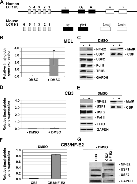
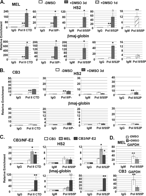
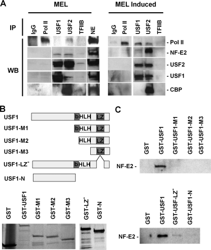
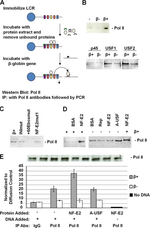
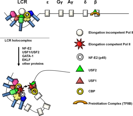
The human beta-globin gene is expressed at high levels in erythroid cells and regulated by proximal and distal cis-acting DNA elements, including promoter, enhancer, and a locus control region (LCR). Transcription complexes are recruited not only to the globin gene promoters but also to the LCR. Previous studies have implicated the ubiquitously expressed transcription factor USF and the tissue-restricted activator NF-E2 in the recruitment of transcription complexes to the beta-globin gene locus. Here we demonstrate that although USF is required for the efficient association of RNA polymerase II (Pol II) with immobilized LCR templates, USF and NF-E2 together regulate the association of Pol II with the adult beta-globin gene promoter. Recruitment of Pol II to the LCR occurs in undifferentiated murine erythroleukemia cells, but phosphorylation of LCR-associated Pol II at serine 5 of the C-terminal domain is mediated by erythroid differentiation and requires the activity of NF-E2. Furthermore, we provide evidence showing that USF interacts with NF-E2 in erythroid cells. The data provide mechanistic insight into how ubiquitous and tissue-restricted transcription factors cooperate to regulate the recruitment and activity of transcription complexes in a tissue-specific chromatin domain.


Figures

FIGURE 1.
FIGURE 1.
DMSO mediated increase in USF, NF-E2 (p45), and β-globin expression in MEL but not CB3 cells. A, structure of the human and mouse β-globin gene loci. The β-globin gene loci consist of five (human) or four (mouse) genes that are expressed during development. The murine ϵγ- and βh1-globin genes are expressed in embryonic erythroid cells, and the βmin- and βmaj-globin genes are expressed in fetal and adult erythroid cells (70). The human ϵ-globin gene is expressed at the embryonic stage, the Gγ- and Aγ-globin genes are expressed at the fetal stage, and the δ- and β-globin genes are expressed at the adult stage. All of the genes are regulated by an LCR located far upstream of the genes and composed of several erythroid-specific HS sites. B, quantitative RT-PCR analysis of βmaj-globin gene expression in MEL cells incubated with or without 1.5% DMSO for 3 days. RNA was isolated from MEL cells, reverse transcribed, and subjected to qPCR using primers specific for the adult βmaj-globin gene. The results are shown as the relative expression normalized to transcription of the β-actin gene. Error bars, S.E. from three independent experiments. C, Western blotting analysis of NF-E2 (p45), USF1, USF2, Pol II, TFIIB, GAPDH, MafK, and CBP in MEL cells incubated with or without 1.5% DMSO for 3 days. Proteins from whole cell extracts (60 μg) were electrophoresed in 4–20% Ready Gels (Bio-Rad), transferred to nitrocellulose membranes, and incubated with antibodies as indicated. D, quantitative RT-PCR analysis of βmaj-globin gene expression in CB3 cells incubated with or without 1.5% DMSO for 3 days. RNA was isolated and analyzed as described in B. E, Western blotting analysis of NF-E2 (p45), USF1, USF2, Pol II, TFIIB, GAPDH, MafK, and CBP in CB3 cells incubated with or without 1.5% DMSO for 3 days. Proteins were processed and analyzed as described in C. F, quantitative RT-PCR analysis of βmaj-globin gene expression in CB3 and CB3/NF-E2 cells. RNA was processed and analyzed as described in B. G, Western blotting analysis of NF-E2 (p45), USF1, USF2, and GAPDH expression in CB3 and CB3/NF-E2 cells. Proteins were processed and analyzed as described in C.
FIGURE 2.
FIGURE 2.
Lack of NF-E2 (p45) impairs the assembly of protein complexes at LCR HS2 and at the adult βmaj-globin gene promoter. A, ChIP analysis of protein chromatin interactions in LCR HS2 and the adult βmaj-globin gene promoter in MEL cells incubated with or without 1.5% DMSO for 3 days. After cross-linking MEL cells with 1% formaldehyde, chromatin was isolated, fragmented by sonication, and subjected to immunoprecipitation with antibodies against NF-E2 (p45), MafK, USF1, USF2, CBP, and TFIIB. Reactions with the IgG antibody served as a negative control. The DNA was purified from the precipitate and subjected to qPCR using primers specific for LCR HS2 and the adult βmaj-globin gene promoter as indicated. Error bars, S.E. of three independent experiments (**, sample versus IgG, p < 0.05; ΔΔ, sample versus IgG, 0.05 < p < 0.1; *, DMSO versus no DMSO, p < 0.05; Δ, DMSO versus no DMSO, 0.05 < p < 0.1). B, ChIP analysis of protein chromatin interactions in LCR HS2 and the adult βmaj-globin gene promoter in CB3 cells incubated with or without 1.5% DMSO for 3 days. DNA was isolated from immunoprecipitated material and analyzed as described in A. Error bars, S.E. of three independent experiments (symbols are as in A). C, comparative ChIP analysis of protein chromatin interactions in uninduced CB3, MEL, and CB3/NF-E2 cells. Cross-linked chromatin was precipitated with IgG or antibodies against NF-E2 (p45), USF1, or USF2, and DNA was analyzed as described in A. Error bars, S.E. of three independent experiments (*, CB3/NF-E2 versus CB3, p < 0.05; Δ, CB3/NF-E2 versus CB3, 0.05 < p < 0.1; **, CB3/NF-E2 versus MEL, p < 0.05; ΔΔ, CB3/NF-E2 versus MEL, 0.05 < p < 0.1; ***, CB3/NF-E2 versus CB3 and MEL p < 0.05).
FIGURE 3.
FIGURE 3.
Efficient recruitment and phosphorylation of Pol II at the β-globin gene locus requires NF-E2 (p45). A, ChIP analysis of Pol II interactions in the β-globin gene locus in MEL cells incubated with or without 1.5% DMSO for 1 or 3 days. Chromatin was isolated from cross-linked MEL cells, fragmented by sonication, and immunoprecipitated with antibodies specific for the Pol II CTD (Pol II/CTD), for unphosphorylated Pol II (Pol II/P), or for Pol II phosphorylated at serine 5 (Pol II/S5P) or serine 2 (Pol II/S2P) of the CTD. IgG or IgM antibodies were used in these experiments as negative controls. DNA was isolated from the precipitates and subjected to qPCR with DNA primers specific for LCR HS2 or the adult βmaj-globin gene promoter as indicated. Error bars, S.E. from three independent experiments (**, sample versus IgG, p < 0.05; *, DMSO versus no DMSO, p < 0.05; Δ, DMSO versus no DMSO, 0.05 < p < 0.1). B, ChIP analysis of Pol II interactions in the β-globin gene locus in CB3 cells incubated with or without 1.5% DMSO for 3 days. Chromatin precipitation and DNA analysis by qPCR was performed as described in A. Error bars, S.E. from three independent experiments (**, as described in A). C, comparative ChIP analysis of Pol II interactions in uninduced CB3, MEL, and CB3/NF-E2 cells. Chromatin precipitation and DNA analysis by qPCR were performed as described in A. Error bars, S.E. from three independent experiments (*, CB3/NF-E2 versus CB3, p < 0.05; **, CB3/NF-E2 versus CB3 and MEL, p < 0.05; ΔΔ, CB3/NF-E2 versus CB3 and MEL, 0.05 < p < 0.1). D, ChIP analysis of Ser-5-phosphorylated Pol II at the GAPDH promoter in undifferentiated and DMSO-induced MEL and CB3 cells (as indicated). Cells were grown in the absence or presence of DMSO (1.5% for 3 days). DNA was isolated from immunoprecipitated material and analyzed by qPCR as described in A. Error bars, S.E. from three independent experiments (**, as described in A).
FIGURE 4.
FIGURE 4.
Interactions of USF1 and USF2 with NF-E2 (p45) and Pol II during differentiation of MEL cells. A, co-immunoprecipitation experiments were performed by first subjecting nuclear extracts from MEL cells incubated with or without 1.5% DMSO for 3 days to immunoprecipitation with antibodies specific for IgG, Pol II (N-20), USF1, USF2, and TFIIB. The immunoprecipitated material was electrophoresed using 4–20% Ready Gels (Bio-Rad) and transferred to nitrocellulose membranes. The nitrocellulose membranes were incubated with antibodies against Pol II, NF-E2 (p45), USF1, USF2, and CBP, as indicated, and subjected to ECL Plus chemiluminescence (Amersham Biosciences). B, generation and expression of USF1/GST fusion proteins in E. coli. The cDNA encoding full-length or truncated USF1 was ligated into the pGEX-5X-1 vector. Fusion proteins were expressed in and purified from E. coli and analyzed by SDS-PAGE. The following USF1-derived proteins were purified: full-length USF1 protein (USF1); deletion of the N-terminal half (USF-M1); deletion of the N terminus and the basic region (USF1-M2); deletion of the N terminus, the basic region, and the helix-loop-helix domain (M3); deletion of the leucine zipper (USF-1 LZ); and deletion of the C terminus as well as the basic region, the HLH domain, and the LZ domain (USF1-N). C, interaction of USF1 with NF-E2. Equal amounts of GST fusion proteins were incubated with His-tagged NF-E2. After washing, proteins were eluted from the GST-beads, electrophoresed using SDS-PAGE, and subjected to Western blotting analysis using NF-E2 (p45)-specific antibodies. WB, Western blot.
FIGURE 5.
FIGURE 5.
USF and NF-E2 regulate the recruitment and dissociation of Pol II to and from immobilized LCR templates. A, scheme of the experimental strategy. A linearized and biotinylated plasmid containing the human β-globin LCR was immobilized on streptavidin-coated magnetic beads as described by Vieira et al. (45). The LCR was then incubated with whole cell extracts from MEL cells. Unbound material was removed, and the LCR-protein complex was washed several times and incubated with different DNA templates in the presence or absence of recombinant NF-E2 (p45) tethered to MafG (16, 42) or dominant negative USF (A-USF) (13). Proteins that dissociate from the LCR after the incubation step were analyzed using Western blotting analysis. Transfer of proteins to the β-globin gene promoter was analyzed by immunoprecipitation (IP) followed by quantitative PCR. B, β-globin promoter-mediated dissociation of Pol II, NF-E2 (p45), USF1, and USF2, from the LCR in the presence of a plasmid containing the β-globin gene with (β+) or without (β) its promoter. Proteins and DNA were removed from the immobilized LCR after incubation for 30 min at 30 °C. C, analysis of the effect of β-globin promoter mutations on the dissociation of Pol II. DNA plasmids containing the wild-type β-globin promoter (β+) or the promoter with mutations in the initiator (INImut), the +60 E-box (+60Eboxmut), or the partial MARE sequence (NF-E2mut1) were incubated for 30 min with the immobilized LCR-protein complex in the presence of recombinant NF-E2. Dissociated proteins were removed and analyzed by Western blotting experiments using an antibody specific for Pol II. D, effect of NF-E2 (p45) and A-USF on the dissociation of Pol II from the immobilized LCR. The immobilized LCR-protein complex was incubated for 30 min with BSA, AAV Rep (Rep), NF-E2, or A-USF in the absence or presence of a plasmid containing the wild-type β-globin gene promoter (β+). Dissociated proteins were removed from the LCR and analyzed by Western blotting experiments using an antibody specific for Pol II. E, quantitative PCR analysis of Pol II transfer from immobilized LCR templates to the β-globin gene promoter. Immobilized LCR-protein complexes were incubated with or without a plasmid containing the β-globin gene with (β+) or without (β) its promoter region for 30 min at 30 °C. Unbound material was subjected to immunoprecipitation using IgG- or Pol II-specific antibodies. The DNA was isolated from the precipitate and subjected to quantitative real-time PCR using primers specific for the β-globin gene. The experiment was repeated, and the error bars represent S.E. An aliquot was taken from immobilized LCR-protein complexes in each transfer dissociation assay and analyzed by Western blotting using Pol II-specific antibodies (shown above the graph).
FIGURE 6.
FIGURE 6.
Model of NF-E2 and USF mediated assembly and transfer of elongation competent transcription complexes in the β-globin gene locus. Incomplete elongation-incompetent Pol II transcription complexes are first recruited to the LCR. This is mediated in part by USF2, its associated co-factor CBP, TFIIB, and other proteins. After erythroid differentiation expression of NF-E2 (p45) and USF increases and these proteins efficiently associate with the LCR. This leads to the recruitment or assembly of transcriptionally competent Pol II complexes and phosphorylation of the Pol II CTD. The differentiation of erythroid cells is also accompanied by a conformational change in the globin locus that juxtaposes the adult globin gene with the LCR. This facilitates the transfer of elongation-competent transcription complexes from the LCR to the adult globin gene.

Similar articles

Cited by

References

    1. Felsenfeld G., Groudine M. (2003) Nature 421, 448–453 - PubMed
    1. Chakalova L., Debrand E., Mitchell J. A., Osborne C. S., Fraser P. (2005) Nat. Rev. Genet. 6, 669–677 - PubMed
    1. Sutherland H., Bickmore W. A. (2009) Nat. Rev. Genet. 10, 457–466 - PubMed
    1. Loose M., Swiers G., Patient R. (2007) Curr. Opin. Hematol. 14, 307–314 - PubMed
    1. Stamatoyannopoulos G. (2005) Exp. Hematol. 33, 259–271 - PMC - PubMed

Publication types

MeSH terms

LinkOut - more resources