. 2010 May 21;285(21):16302-14.
doi: 10.1074/jbc.M109.072439. Epub 2010 Mar 17.

Sphingosine 1-phosphate and sphingosine kinase are involved in a novel signaling pathway leading to acrosomal exocytosis

Affiliations

Sphingosine 1-phosphate and sphingosine kinase are involved in a novel signaling pathway leading to acrosomal exocytosis

Laila Suhaiman et al. J Biol Chem. .

Abstract

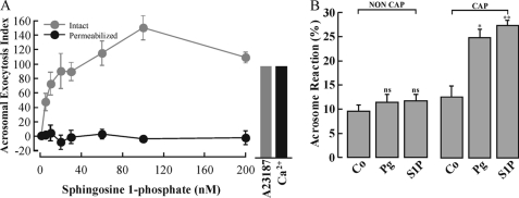
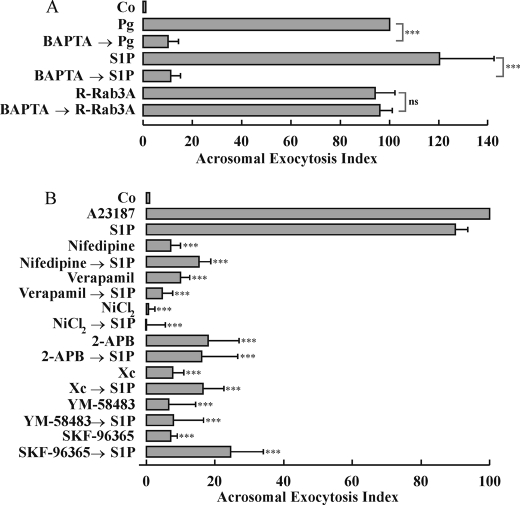
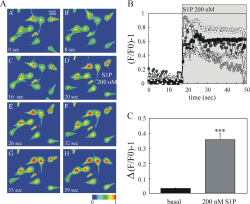
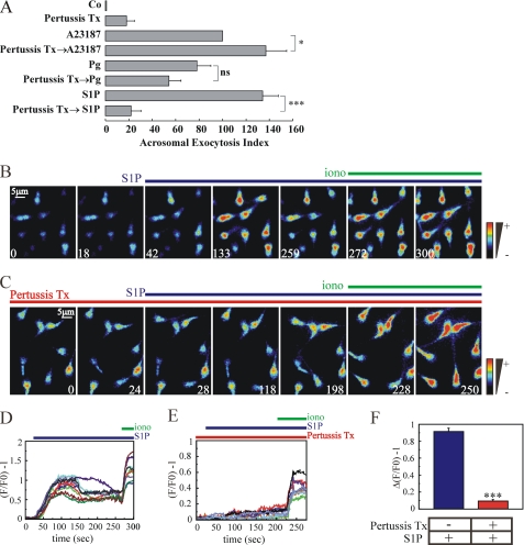
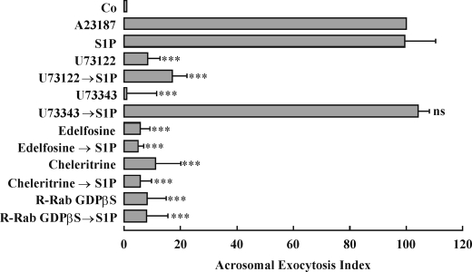
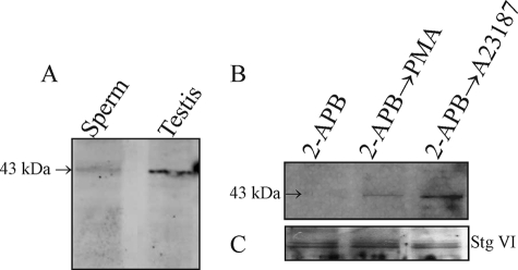
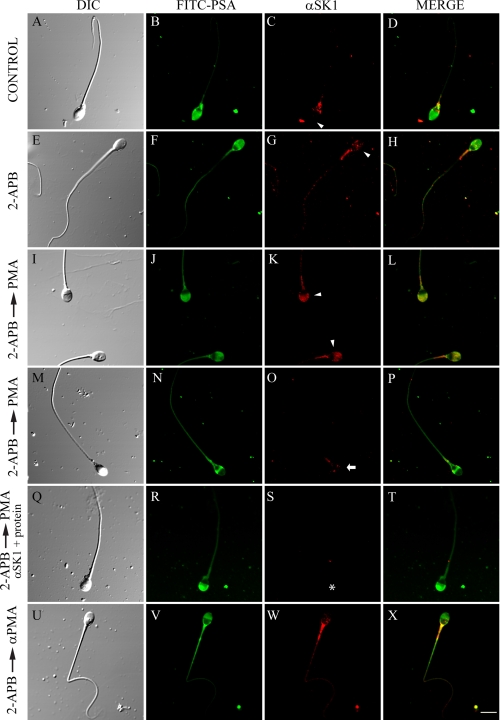
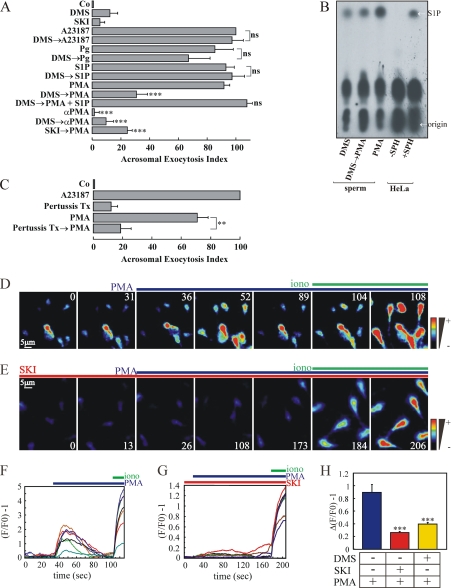
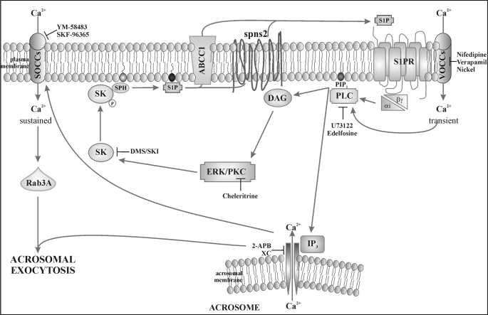
Regulated secretion is a central issue for the specific function of many cells; for instance, mammalian sperm acrosomal exocytosis is essential for egg fertilization. Sphingosine 1-phosphate is a bioactive sphingolipid that regulates crucial physiological processes. Here we report that this lipid triggers acrosomal exocytosis in human sperm by a mechanism involving a G(i)-coupled receptor. Real-time imaging showed a remarkable increase of cytosolic calcium upon activation with sphingosine 1-phosphate and pharmacological experiments indicate that the process requires extracellular calcium influx through voltage and store-operated calcium channels and efflux from intracellular stores through inositol 1,4,5-trisphosphate-sensitive calcium channels. Sphingosine 1-phosphate-induced exocytosis requires phospholipase C and protein kinase C activation. We investigated possible sources of the lipid. Western blot indicates that sphingosine kinase 1 is present in spermatozoa. Indirect immunofluorescence showed that phorbol ester, a potent protein kinase C activator that can also trigger acrosomal exocytosis, redistributes sphingosine kinase 1 to the acrosomal region. Functional assays showed that phorbol ester-induced exocytosis depends on the activation of sphingosine kinase 1. Furthermore, incorporation of (32)P to sphingosine demonstrates that cells treated with the phorbol ester increase their sphingosine kinase activity that yields sphingosine 1-phosphate. We present here the first evidence indicating that human spermatozoa produce sphingosine 1-phosphate when challenged with an exocytic stimulus. These observations point to a new role of sphingosine 1-phosphate in a signaling cascade that facilitates acrosome reaction providing some clues about novel lipid molecules involved in exocytosis.


Figures

FIGURE 1.
FIGURE 1.
S1P triggers acrosomal exocytosis in intact human spermatozoa. A, after swim-up in HTF (5 mg/ml bovine serum albumin) at 37°C, 5% CO2, sperm were incubated for an additional 2 h under capacitating conditions. Intact and streptolysin O-permeabilized sperm were used. Sperm were treated with increasing concentrations of S1P (0–200 nm) for 15 min at 37°C in 5% CO2. As positive control, permeabilized spermatozoa were stimulated with 10 μm free Ca2+ (black bar) and intact sperm with 10 μm A23187 (gray bar). Sperm were fixed and acrosomal exocytosis was evaluated by FITC-PSA binding with at least 300 cells per condition scored. The data represent the mean ± S.E. from three to five independent experiments. B, an aliquot of a sperm sample was subjected to swim-up in HTF medium under non-capacitating conditions (37°C 5% CO2, without bovine serum albumin). Another aliquot was processed in capacitating conditions: swim-up in HTF (5 mg/ml bovine serum albumin, 37°C, 5% CO2). Cells recovered from swim-up were incubated for an additional 2 h under non-capacitating or capacitating conditions. Sperm were treated or not (control, Co) with 15 μm progesterone (Pg) or 100 nm S1P for 15 min at 37°C 5% CO2. Acrosomal exocytosis was evaluated as explained under “Experimental Procedures.” The data represent the mean ± S.E. of at least four independent experiments. The means of groups NON CAP and CAP were compared with the corresponding control using Dunnett's test and classified as non-significant (ns, p > 0.05), or significant (*, p < 0.01 or **, p < 0.001).
FIGURE 2.
FIGURE 2.
S1P-induced exocytosis requires extracellular calcium influx and acrosomal calcium efflux. A, capacitated spermatozoa were incubated at 37 °C for 15 min without any stimulus (control, Co) or treated with 15 μm progesterone (Pg), 100 nm S1P, or 1 μm cell-permeant prenylated Rab3A activated with GTPγS (R-Rab3A). When indicated, cells were treated before the stimulus with 5 mm BAPTA (BAPTAPg, BAPTAS1P, and BAPTAR-Rab3A) at 37 °C for 15 min. The data represent the mean ± S.E. of at least four independent experiments. The means of groups with BAPTA were compared with the corresponding group without the chelator using Student's t-test and classified as significant (***, p < 0.001) or non-significant (ns, p > 0.05). B, capacitated sperm were incubated without any treatment (control, Co) or treated with 10 μm nifedipine, 100 μm verapamil, 0.1 mm NiCl2, 1 μm YM-58483, 50 μm SKF-96365, 100 μm 2-APB, or 1.1 μm xestospongin C (Xc) for 15 min at 37 °C. When specified, acrosomal exocytosis was activated by adding 100 nm S1P, and the incubation continued for an additional 15 min (NifedipineS1P, VerapamilS1P, NiCl2S1P, YM-58483S1P, SKF-96365S1P, 2-APBS1P, and XcS1P). A control with the Ca2+ ionophore A23187 (10 μm) was included. Sperm were then fixed, and acrosomal exocytosis was measured as described under “Experimental Procedures.” The data represent the mean ± S.E. from 3 to 14 independent experiments and were normalized as described under “Experimental Procedures.” Dunnett's test was used to compare the means of all groups against the S1P-stimulated condition in the absence of inhibitors. Significant differences at p < 0.001(***) are indicated for each bar.
FIGURE 3.
FIGURE 3.
S1P causes a [Ca2+]i increase in human sperm. Capacitated human sperm recovered after swim-up were loaded with Fluo-3 AM (2 μm) in HSM. The fluorescence intensity was visualized before and after S1P addition as described under “Experimental Procedures.” A, representative images of Fluo-3 AM-loaded human sperm before (0, 8, and 16 s) and after (20, 26, 32, 53, and 59 s) the application of 200 nm S1P. The color bar shows fluorescence intensity after background subtraction. B, representative traces of five individual human sperm. The plots indicate the increase in fluorescence intensity in response to 200 nm S1P addition. Fluorescence is expressed as (F/F0) − 1 versus time. C, the increase of fluorescence upon stimulation is shown as the average of all individual human sperm analyzed (n = 101). Error bars represent the mean ± S.E.. Note: approximately 45% of the cells responded to S1P addition with an intracellular calcium increase.
FIGURE 4.
FIGURE 4.
S1P triggers exocytosis through a Gi-coupled receptor. A, intact capacitated human sperm were incubated with or without 100 ng/ml pertussis toxin (Pertussis Tx) for 15 min at 37°C. Acrosomal exocytosis was then initiated by adding 10 μm A23187 (Pertussis TxA23187), 15 μm progesterone (Pertussis TxPg), or 100 nm S1P (Pertussis TxS1P) for a further 15 min at 37°C. Several controls were included: background acrosomal exocytosis in the absence of any stimulation (control, Co); acrosomal exocytosis stimulated by 10 μm A23187 (A23187), 15 μm progesterone (Pg), or 100 nm S1P (S1P). Afterwards, sperm were fixed, and acrosomal exocytosis was measured as described under “Experimental Procedures.” The data represent the mean ± S.E. from 4 to 8 independent experiments. The means of groups with toxin were compared with the corresponding group without the inhibitor using Student's t-test and classified as significant (*, p < 0.05; ***, p < 0.001) or non-significant (ns, p > 0.05). B–F, capacitated human sperm recovered after swim-up were loaded with Fluo-3 AM (2 μm) in HSM, and the fluorescence intensity was visualized before and after S1P addition as described under “Experimental Procedures.” B and C, representative single cell spatiotemporal [Ca2+]i changes after adding 200 nm S1P (blue line) in the absence (B) or presence (C) of 100 ng/ml pertussis toxin (Pertussis Tx-red line). Ionomycin (20 μm iono, green line) was added at end the of the experiment as a positive control. The time frame is indicated in each panel (seconds). D and E, illustrate the corresponding traces of individual sperm showing the fluorescence change after addition of S1P in absence (D) or presence (E) of pertussis toxin. F, summarizes the S1P response in the absence (blue bar) or presence of 100 ng/ml pertussis toxin (red bar). Δ(F/F0) − 1 represents the average of the changes in fluorescence of all individual human sperm analyzed (S1P (N = 7, 145 cells) and Pertussis Tx+S1P (N = 4, 96 cells), *** (p <=0.001)). Error bars represent the mean ± S.E. Note: approximately 45% of the cells responded to S1P addition with an intracellular calcium increase.
FIGURE 5.
FIGURE 5.
S1P-induced acrosomal exocytosis requires PLC, PKC activities, and Rab3A. Human spermatozoa were incubated for 3 h under capacitating conditions. The medium was then supplemented, as indicated, with the following compounds: 15 μm U73122, 15 μm U73343 (inactive analogue of U73122), 10 μm cheleritrine, or 1 μm R-Rab3A-GDPβS (unprenylated cell-permeant Rab3A, loaded with GDPβS). The samples were further incubated for 15 min at 37 °C with no addition (Co) or in the presence of 100 nm S1P (U73122S1P, U73343S1P, CheleritrineS1P, and R-Rab3A-GDPβSS1P). As positive control 10 μm A23187 was used. Acrosomal exocytosis was evaluated as explained under “Experimental Procedures.” The data represent the mean ± S.E. from three to eleven independent experiments. Dunnett's test was used to compare the means of all groups against the S1P-stimulated condition in the absence of inhibitors. Significant, p < 0.001(***) or non-significant (ns, p > 0.05) differences are indicated for each bar.
FIGURE 6.
FIGURE 6.
Sphingosine kinase 1 is present in human sperm. A, whole sperm proteins were extracted in Laemmli sample buffer (10 × 106 cells) and analyzed by Western blot with a rabbit polyclonal anti-SK1 antibody (Sperm). A testis extract (Testis) was used as a control. Molecular mass standards (kDa) are indicated on the right. B, sperm incubated for 3 h under capacitating conditions were treated with 100 μm 2-APB for 15 min (2-APB), two samples were further incubated for 15 min at 37°C with 200 nm PMA or 10 μm A23187 (2-APBPMA and 2-APBA23187). After treatment, membranes from each sample (50 × 106 cells) were obtained according to Bohring and Krause (23). The samples were analyzed by Western blot using the anti-SK1 antibody. C, the blotted Immobilon membranes were stripped and reprobed with an anti-synaptotagmin VI antibody. Shown is a blot representative of three experiments.
FIGURE 7.
FIGURE 7.
Sphingosine kinase 1 translocates from the postacrosomal to the acrosomal region in stimulated human spermatozoa. Capacitated human sperm were incubated or not with 100 μm 2-APB (15 min at 37 °C) as explained in the legend to Fig. 6B (A, E, I, M, Q, and U). The cells were then fixed and double-stained with an anti-SK1 antibody followed by an anti-rabbit Cy3 (red: C, G, K, O, S, and W) and FITC-PSA to differentiate between reacted and intact sperm (green: B, F, J, N, R, and V). The merged images are shown in D, H, L, P, T, and X. Some batches were further incubated with 200 nm PMA (2-APBPMA) or its inactive analogue αPMA (2-APB→αPMA). As an antibody-specificity control 10 μg/ml of the anti-SK1 antibody was preincubated with 0.6 μg/ml of the SK1 protein (2-APBPMA, αSK1+protein, asterisk). Bar = 6 μm.
FIGURE 8.
FIGURE 8.
SK1 activity is required for the PMA-triggered signal transduction cascade leading to acrosomal exocytosis. A, human spermatozoa were incubated for 3 h under capacitating conditions. The medium was then supplemented, when indicated, with 5 μm DMS or 1 μm SKI. Acrosomal exocytosis was then initiated by adding 10 μm A23187 (DMSA23187), 15 μm progesterone (DMSPg), 100 nm S1P (DMSS1P), 200 nm PMA (DMSPMA and SKIPMA), 200 nm PMA plus 100 nm S1P (DMSPMA + S1P), or αPMA (inactive analogue of PMA; DMS→αPMA). The mixture was incubated for 15 min at 37 °C. Several controls were included: background acrosomal exocytosis in the absence of any stimulation (control, Co); acrosomal exocytosis stimulated by 10 μm A23187 (A23187), 15 μm progesterone (Pg), 100 nm S1P (S1P), 200 nm PMA (PMA), or 200 nm αPMA (αPMA). Afterwards, sperm were fixed and acrosomal exocytosis was measured as described under “Experimental Procedures.” The data represent the mean ± S.E. of at least four independent experiments. In the case of stimulation with A23187, Pg, or S1P, the means of groups with DMS were compared with the corresponding group without the inhibitor using the Student's t-test; when PMA or αPMA were used as stimulators, the Dunnett's test was used to compare the means of all groups against the PMA-stimulated condition in the absence of inhibitors (ns, p > 0.05; s, p < 0.001 (***)). B, PMA increases S1P synthesis in human sperm cells. Human spermatozoa (50 × 106) were permeabilized (as described under “Experimental Procedures”) and treated for 15 min at 37 °C with 5 μm DMS. Samples were further incubated for 15 min at 37 °C with 50 μm bovine serum albumin-sphingosine, 1 μl of [γ-32P]ATP, and when indicated 200 nm PMA (DMSPMA and PMA). HeLa cells (positive control, incubated with sphingosine; +SPH, and negative control no SPH added; −SPH) were prepared as described under “Experimental Procedures.” Thin-layer chromatograms were developed in 1-butanol/methanol/acetic acid/water (8:2:1:2, v/v) as solvent system and visualized by autoradiography. The figure is representative of three experiments. C, intact capacitated human sperm were incubated with or without 100 ng/ml pertussis toxin (Pertussis Tx) for 15 min at 37 °C. Acrosomal exocytosis was then initiated by adding 200 nm PMA (Pertussis TxPMA) and incubating for 15 min at 37 °C. Several controls were included: background acrosomal exocytosis in the absence of any stimulation (control, Co); acrosomal exocytosis stimulated by 10 μm A23187 (A23187) or 200 nm PMA (PMA). Afterwards, sperm were fixed and acrosomal exocytosis was measured as described under “Experimental Procedures.” The data represent the mean ± S.E. of five independent experiments. The mean of the group with toxin was compared with the corresponding group without the inhibitor using Student's t-test and classified as significant (**, p < 0.01). D–H, capacitated human sperm recovered after swim up were loaded with Fluo-3 AM (2 μm) in HSM, and the fluorescence intensity was visualized before and after PMA addition as described under “Experimental Procedures.” D and E, representative single cell spatiotemporal [Ca2+]i changes and their corresponding traces after adding 200 nm PMA (blue line) in the absence (D) or presence (E) of 1 μm SKI (SKI-red line). Ionomycin (20 μm, iono, green line) was added at the end of the experiment as positive control. The time frame is indicated in each panel (seconds). F and G, illustrate the corresponding traces of individual sperm showing the fluorescence change after addition of PMA in the absence (F) or presence (G) of SKI. H, summarizes the PMA response in the absence (blue bar) or presence of 1 μm SKI (red bar) or 5 μm of DMS (yellow bar). Δ(F/F0) − 1 is the average of the changes in fluorescence of all individual human sperm analyzed (PMA (N = 8, 135 cells), SKI+PMA (N = 5, 67 cells), and DMS+PMA (N = 4, 53 cells)). Error bars represent the mean ± S.E. Dunnett's test was used to compare the means of all groups against the PMA-stimulated condition in the absence of inhibitors. Significant differences from PMA group for p < 0.001 (***) are indicated for each bar.
FIGURE 9.
FIGURE 9.
Working model for S1P/SK/S1PR interplay during acrosomal exocytosis. The interaction between S1P with a Gi-coupled receptor (S1PR) activates a VOCC and a heterotrimeric Gi-protein leading to PLC activation. PLC hydrolyzes phosphatidylinositol 4,5-bisphosphate to generate DAG and IP3. IP3 binds to IP3-sensitive calcium channels on the acrosomal membrane leading to intra-acrosomal calcium efflux. The emptying of the acrosomal store stimulates the opening of SOCCs at the plasma membrane. The other product of PLC activity, DAG, activates the serine/threonine kinase PKC. PKC may act directly or indirectly through ERK phosphorylating SK1 on residue Ser225 and favoring its interaction with the plasma membrane. Once the enzyme resides on the membrane, it catalyzes sphingosine (SPH) phosphorylation. S1P produced reaches the extracellular medium through an ABCC1 transporter and/or a Spns2 transporter interacting with its receptor and amplifying the signaling pathway. The sustained calcium increase induced by SOCCs opening activates Rab3A leading to acrosomal exocytosis.

Similar articles

Cited by

References

    1. Spiegel S., English D., Milstien S. (2002) Trends Cell Biol. 12, 236–242 - PubMed
    1. Taha T. A., Argraves K. M., Obeid L. M. (2004) Biochim. Biophys. Acta 1682, 48–55 - PubMed
    1. Yatomi Y. (2006) Curr. Pharm. Des. 12, 575–587 - PubMed
    1. Rosen H., Goetzl E. J. (2005) Nat. Rev. Immunol. 5, 560–570 - PubMed
    1. Sanchez T., Hla T. (2004) J. Cell Biochem. 92, 913–922 - PubMed

Publication types

MeSH terms

LinkOut - more resources