. 2010 May;153(1):337-47.
doi: 10.1104/pp.110.154690. Epub 2010 Mar 17.

RNA interference-mediated change in protein body morphology and seed opacity through loss of different zein proteins

Affiliations

RNA interference-mediated change in protein body morphology and seed opacity through loss of different zein proteins

Yongrui Wu et al. Plant Physiol. 2010 May.

Abstract

Opaque or nonvitreous phenotypes relate to the seed architecture of maize (Zea mays) and are linked to loci that control the accumulation and proper deposition of storage proteins, called zeins, into specialized organelles in the endosperm, called protein bodies. However, in the absence of null mutants of each type of zein (i.e. alpha, beta, gamma, and delta), the molecular contribution of these proteins to seed architecture remains unclear. Here, a double null mutant for the delta-zeins, the 22-kD alpha-zein, the beta-zein, and the gamma-zein RNA interference (RNAi; designated as z1CRNAi, betaRNAi, and gammaRNAi, respectively) and their combinations have been examined. While the delta-zein double null mutant had negligible effects on protein body formation, the betaRNAi and gammaRNAi alone only cause slight changes. Substantial loss of the 22-kD alpha-zeins by z1CRNAi resulted in protein body budding structures, indicating that a sufficient amount of the 22-kD zeins is necessary for maintenance of a normal protein body shape. Among different mutant combinations, only the combined betaRNAi and gammaRNAi resulted in drastic morphological changes, while other combinations did not. Overexpression of alpha-kafirins, the homologues of the maize 22-kD alpha-zeins in sorghum (Sorghum bicolor), in the beta/gammaRNAi mutant failed to offset the morphological alterations, indicating that beta- and gamma-zeins have redundant and unique functions in the stabilization of protein bodies. Indeed, opacity of the beta/gammaRNAi mutant was caused by incomplete embedding of the starch granules rather than by reducing the vitreous zone.


Figures

Figure 1.
Figure 1.
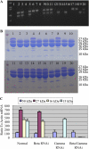
Zein accumulation in normal and RNAi mutant seeds detected by SDS-PAGE and PCR. A, SDS-PAGE of 12 different maize inbred lines and genetic varieties. Lane numbers refer to different materials: 1, BSSS53; 2, B73; 3, B37; 4, Mo17; 5, W64A; 6, W22; 7, P1-ww-112; 8, A69Y; 9, ILLIZE; 10, A188; 11, SD-purple; 12, A654. Bands for 27-kD γ, 22-kD α, 19-kD α, 16-kD γ, 15-kD β, and 10-kD δ are well separated. Several lines are missing the 10-kD δ-zein. B, The βRNAi mutant. The top panel shows the construct (see “Materials and Methods”). The middle panel shows PCR assay of genomic DNA from different transgenic lines. K1, K4, and K8 represent the progeny inheriting the RNAi event. Two nontransgenic kernels, C1 and C2, serve as controls. The bottom panel shows the corresponding SDS-PAGE. In lanes K1, K4, and K8, the 15-kD band is missing. C, The γRNAi mutant. Analysis is the same as in B. K3 and K4 represent the progeny inheriting the RNAi event. D, SDS-PAGE for a z1CRNAi seed is shown, where the 22-kD zein band is reduced (arrow). E, SDS-PAGE for W64A o2 and normal W64A seeds is shown. In the o2 mutant, bands for 22-kD α-zein, 15-kD β-zein, and 10-kD δ-zein are reduced (arrows). Total zein loaded in each lane was equal to 300 μg of dry seed meal (A) and 500 μg of fresh endosperm at 18 DAP (B–D). Protein markers from top to bottom are 25, 20, 15, and 10 kD. M, Protein marker; F and R, primer GFPF and T35S-HindIII (see “Materials and Methods”). [See online article for color version of this figure.]
Figure 2.
Figure 2.
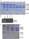
Specificity of RNAi events. Genotypes were identified with PCR (A), protein accumulation with SDS-PAGE (B), and mRNA levels with real-time RT-PCR (C), as described in “Materials and Methods.” Lanes (K1–K20) are numbered for each kernel analyzed by PCR and SDS-PAGE. K2, K3, K5, K9, K10, K11, and K20 inherited both of the RNAi constructs and therefore lack accumulation of βRNAi, the 27- and 16-kD γ-zeins. K1, K4, K8, K12, K14, and K16 were segregants, showing normal protein accumulation. The rest of the individuals inherited one or the other RNAi event, lacking only the corresponding protein. C, For mRNA levels, the endosperms with the same genotype were combined. Their RNAs were extracted for real-time PCR. Error bars indicate sd of three replicates. Total zein loaded in each lane was equal to 500 μg of fresh endosperm at 18 DAP. Protein markers from top to bottom are 50, 20, and 15 kD. [See online article for color version of this figure.]
Figure 3.
Figure 3.
Ultrastructural protein body morphologies of different genotypes. A, BA normal type. B, Inbred A654 (δ-zein double null mutant). C, The βRNAi mutant. D, The γRNAi mutant. E, W64A o2. F, The z1CRNAi mutant. Protruding protein bodies in the z1CRNAi mutant are marked with arrows. Mt, Mitochondria; Pb, protein body; SG, starch granule. Bars = 500 nm.
Figure 4.
Figure 4.
Combinations created by cross or back-cross. A, SDS-PAGE of total zeins from seeds of different crosses is shown; lanes with different backgrounds are labeled, and band sizes are indicated. B, Combinations with A654. A specific primer pair for the δ-zein genes detects the absence of either gene as shown in the BA control. C, Heterologous expression of the 22-kD kafirin genes. SDS-PAGE of seeds from the kafirin transgenic plant and its combination with the βRNAi and γRNAi is shown. Total zein loaded in each lane was equal to 500 μg of fresh endosperm at 18 DAP. Protein markers from top to bottom are 25, 20, 15, and 10 kD. [See online article for color version of this figure.]
Figure 5.
Figure 5.
Ultrastructural protein body morphologies of a series of mutant combinations. A, Combination of the δ-zein double null mutant and βRNAi. B, Combination of the δ-zein double null mutant and γRNAi. C, Combination of the βRNAi and γRNAi. D, Combination of the kafirin transgenes and the βRNAi and γRNAi. Pb, Protein body; SG, starch granule. Bars = 500 nm.
Figure 6.
Figure 6.
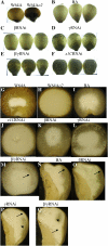
Kernel phenotypes of the βRNAi and γRNAi and their combination. A, BA nontransgenic ear. B, Selfed T2 βRNAi ear. C, T2 homozygous γRNAi ear. D, Cross of a homozygous γRNAi mutant with a heterozygous βRNAi mutant. E, Selfed progeny from D with segregating opaque and vitreous kernels (arrows). F, Genotyping of opaque and vitreous seeds by PCR (see “Materials and Methods”); the 1,096-bp band represents the γRNAi event, and the 913-bp band represents the βRNAi event. G, SDS-PAGE analysis of vitreous and opaque seeds. Band sizes of different zeins are indicated. Total zein loaded in each lane was equal to 300 μg of dry seed meal. Protein markers from top to bottom are 25, 20, 15, and 10 kD. [See online article for color version of this figure.]
Figure 7.
Figure 7.
Kernel opacity of the RNAi mutants. A to F, Translucency of intact kernels on a light box. A, W64A and W64A o2. B, BA normal kernels. C, The βRNAi mutant. D, The γRNAi mutant. E, The βRNAi and γRNAi combination. F, z1CRNAi. G to Q, Latitudinal and longitudinal sections of kernels. Vitreous region was largely reduced in W64A o2 (H) and z1CRNAi (J). In the γRNAi mutant (L and P), the starch granules began to penetrate outside. The crown of the γRNAi mutant seed was opaque (P), while the normal BA kernels were vitreous (N; arrow). Most of the seed trunk of the γRNAi mutant still remained vitreous (P; arrowhead). The penetration was reinforced in the combined mutant of the βRNAi and γRNAi (M and Q), with no reduction of vitreous width (M and Q; arrows). Still, vitreous patches could be seen (Q; arrowhead). G to M show latitudinal sections of W64A (G), W64A o2 (H), BA normal type (I), the z1CRNAi mutant (J), the βRNAi mutant (K), the γRNAi mutant (L), and the βRNAi and γRNAi combination (M). N to Q show longitudinal sections of BA normal type (N), the βRNAi mutant (O), the γRNAi mutant (P), and the βRNAi and γRNAi combination (Q). [See online article for color version of this figure.]
Figure 8.
Figure 8.
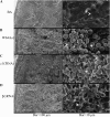
Scanning electron micrographs of the peripheral regions of decapped wild-type and mutant dry kernels. A, BA. B, W64A o2. C, The z1CRNAi mutant. D, The βRNAi and γRNAi combination. The starch granules (arrows) and the proteinaceous matrix mixed with broken protein bodies (arrowheads) are indicated. Bars at left = 100 μm; bars at right = 10 μm.

Similar articles

Cited by

References

    1. Athma P, Peterson T. (1991) Ac induces homologous recombination at the maize P locus. Genetics 128: 163–173 - PMC - PubMed
    1. Bagga S, Adams HP, Rodriguez FD, Kemp JD, Sengupta-Gopalan C. (1997) Coexpression of the maize delta-zein and beta-zein genes results in stable accumulation of delta-zein in endoplasmic reticulum-derived protein bodies formed by beta-zein. Plant Cell 9: 1683–1696 - PMC - PubMed
    1. Burr B, Burr FA. (1976) Zein synthesis in maize endosperm by polyribosomes attached to protein bodies. Proc Natl Acad Sci USA 73: 515–519 - PMC - PubMed
    1. Chandrashekar A, Mazhar H. (1999) The biochemical basis and implications of grain strength in sorghum and maize. J Cereal Sci 30: 193–207
    1. Coleman CE, Clore AM, Ranch JP, Higgins R, Lopes MA, Larkins BA. (1997) Expression of a mutant alpha-zein creates the floury2 phenotype in transgenic maize. Proc Natl Acad Sci USA 94: 7094–7097 - PMC - PubMed

Publication types

LinkOut - more resources