Kinetic analysis of 18F-fluoride PET images of breast cancer bone metastases
- PMID: 20237040
- PMCID: PMC3063528
- DOI: 10.2967/jnumed.109.070052
Kinetic analysis of 18F-fluoride PET images of breast cancer bone metastases
Abstract
The most common site of metastasis for breast cancer is bone. Quantitative (18)F-fluoride PET can estimate the kinetics of fluoride incorporation into bone as a measure of fluoride transport, bone formation, and turnover. The purpose of this analysis was to evaluate the accuracy and precision of (18)F-fluoride model parameter estimates for characterizing regional kinetics in metastases and normal bone in breast cancer patients.
Methods: Twenty metastatic breast cancer patients underwent dynamic (18)F-fluoride PET. Mean activity concentrations were measured from serial blood samples and regions of interest placed over bone metastases, normal vertebrae, and cardiac blood pools. This study examined parameter identifiability, model sensitivity, error, and accuracy using parametric values from the patient cohort.
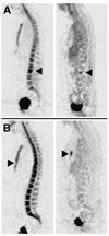
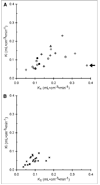
Results: Representative time-activity curves and model parameter ranges were obtained from the patient cohort. Model behavior analyses of these data indicated (18)F-fluoride transport and flux (K(1) and Ki, respectively) into metastatic and normal osseous tissue could be independently estimated with a reasonable bias of 9% or less and reasonable precision (coefficients of variation <or= 16%). Average (18)F-fluoride transport and flux into metastases from 20 patients (K(1) = 0.17 +/- 0.08 mL x cm(-3) x min(-1) and Ki = 0.10 +/- 0.05 mL x cm(-3) x min(-1)) were both significantly higher than for normal bone (K(1) = 0.09 +/- 0.03 mL x cm(-3) x min(-1) and Ki = 0.05 +/- 0.02 mL x cm(-3) x min(-1), P < 0.001).
Conclusion: Fluoride transport and flux can be accurately and independently estimated for bone metastases and normal vertebrae. Reasonable bias and precision for estimates of K(1) and Ki from simulations and significant differences in values from patient modeling results in metastases and normal bone suggest that (18)F-fluoride PET images may be useful for assessing changes in bone turnover in response to therapy. Future studies will examine the correlation of parameters to biologic features of bone metastases and to response to therapy.
Figures


Similar articles
-
Is Response Assessment of Breast Cancer Bone Metastases Better with Measurement of 18F-Fluoride Metabolic Flux Than with Measurement of 18F-Fluoride PET/CT SUV?J Nucl Med. 2019 Mar;60(3):322-327. doi: 10.2967/jnumed.118.208710. Epub 2018 Jul 24. J Nucl Med. 2019. PMID: 30042160 Free PMC article.
-
Castration-resistant prostate cancer bone metastasis response measured by 18F-fluoride PET after treatment with dasatinib and correlation with progression-free survival: results from American College of Radiology Imaging Network 6687.J Nucl Med. 2015 Mar;56(3):354-60. doi: 10.2967/jnumed.114.146936. Epub 2015 Jan 29. J Nucl Med. 2015. PMID: 25635138 Free PMC article. Clinical Trial.
-
A prospective study of risedronate on regional bone metabolism and blood flow at the lumbar spine measured by 18F-fluoride positron emission tomography.J Bone Miner Res. 2003 Dec;18(12):2215-22. doi: 10.1359/jbmr.2003.18.12.2215. J Bone Miner Res. 2003. PMID: 14672357
-
A literature review of 18F-fluoride PET/CT and 18F-choline or 11C-choline PET/CT for detection of bone metastases in patients with prostate cancer.Nucl Med Commun. 2013 Oct;34(10):935-45. doi: 10.1097/MNM.0b013e328364918a. Nucl Med Commun. 2013. PMID: 23903557 Review.
-
Input function and modeling for determining bone metabolic flux using [18 F] sodium fluoride PET imaging: A step-by-step guide.Med Phys. 2023 Apr;50(4):2071-2088. doi: 10.1002/mp.16125. Epub 2022 Dec 17. Med Phys. 2023. PMID: 36433629 Review.
Cited by
-
The isotope bone scan: we can do better.Eur J Nucl Med Mol Imaging. 2013 Aug;40(8):1139-40. doi: 10.1007/s00259-013-2439-2. Epub 2013 May 15. Eur J Nucl Med Mol Imaging. 2013. PMID: 23674209 No abstract available.
-
Can PET-CT imaging and radiokinetic analyses provide useful clinical information on atypical femoral shaft fracture in osteoporotic patients?Curr Osteoporos Rep. 2012 Mar;10(1):42-7. doi: 10.1007/s11914-011-0088-6. Curr Osteoporos Rep. 2012. PMID: 22286527
-
Comparison of standardized uptake values measured on F-NaF PET/CT scans using three different tube current intensities.Radiol Bras. 2015 Jan-Feb;48(1):17-20. doi: 10.1590/0100-3984.2014.0034. Radiol Bras. 2015. PMID: 25798003 Free PMC article.
-
Quantification of skeletal blood flow and fluoride metabolism in rats using PET in a pre-clinical stress fracture model.Mol Imaging Biol. 2012 Jun;14(3):348-54. doi: 10.1007/s11307-011-0505-3. Mol Imaging Biol. 2012. PMID: 21785919 Free PMC article.
-
Prediction of therapy response in bone-predominant metastatic breast cancer: comparison of [18F] fluorodeoxyglucose and [18F]-fluoride PET/CT with whole-body MRI with diffusion-weighted imaging.Eur J Nucl Med Mol Imaging. 2019 Apr;46(4):821-830. doi: 10.1007/s00259-018-4223-9. Epub 2018 Dec 1. Eur J Nucl Med Mol Imaging. 2019. PMID: 30506455 Free PMC article. Clinical Trial.
References
-
- Hamaoka T, Madewell JE, Podoloff DA, Hortobagyi GN, Ueno NT. Bone imaging in metastatic breast cancer. J Clin Oncol. 2004;22:2942–2953. - PubMed
-
- Even-Sapir E. Imaging of malignant bone involvement by morphologic, scintigraphic, and hybrid modalities. J Nucl Med. 2005;46:1356–1367. - PubMed
-
- Petren-Mallmin M, Andreasson I, Ljunggren O, et al. Skeletal metastases from breast cancer: uptake of 18F-fluoride measured with positron emission tomography in correlation with CT. Skeletal Radiol. 1998;27:72–76. - PubMed
-
- Grynpas MD. Fluoride effects on bone crystals. J Bone Miner Res. 1990;5 suppl 1:S169–S175. - PubMed
-
- Hawkins RA, Choi Y, Huang SC, et al. Evaluation of the skeletal kinetics of fluorine-18-fluoride ion with PET. J Nucl Med. 1992;33:633–642. - PubMed
Publication types
MeSH terms
Substances
Grants and funding
LinkOut - more resources
Full Text Sources
Medical
Miscellaneous
