

Kinetic analysis of 18F-fluoride PET images of breast cancer bone metastases
- PMID: 20237040
- PMCID: PMC3063528
- DOI: 10.2967/jnumed.109.070052
Kinetic analysis of 18F-fluoride PET images of breast cancer bone metastases
Abstract
The most common site of metastasis for breast cancer is bone. Quantitative (18)F-fluoride PET can estimate the kinetics of fluoride incorporation into bone as a measure of fluoride transport, bone formation, and turnover. The purpose of this analysis was to evaluate the accuracy and precision of (18)F-fluoride model parameter estimates for characterizing regional kinetics in metastases and normal bone in breast cancer patients.
Methods: Twenty metastatic breast cancer patients underwent dynamic (18)F-fluoride PET. Mean activity concentrations were measured from serial blood samples and regions of interest placed over bone metastases, normal vertebrae, and cardiac blood pools. This study examined parameter identifiability, model sensitivity, error, and accuracy using parametric values from the patient cohort.
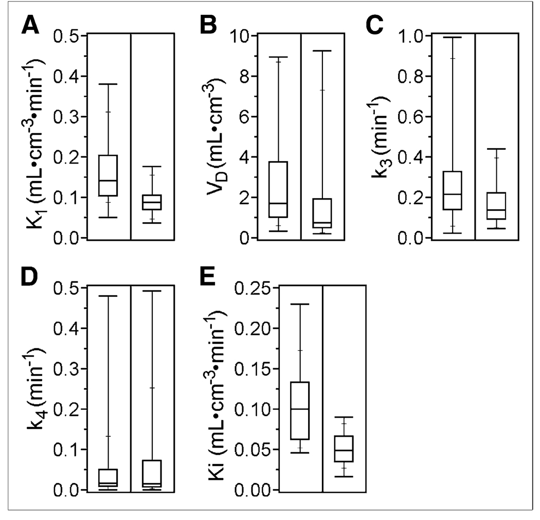
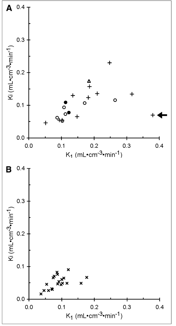
Results: Representative time-activity curves and model parameter ranges were obtained from the patient cohort. Model behavior analyses of these data indicated (18)F-fluoride transport and flux (K(1) and Ki, respectively) into metastatic and normal osseous tissue could be independently estimated with a reasonable bias of 9% or less and reasonable precision (coefficients of variation <or= 16%). Average (18)F-fluoride transport and flux into metastases from 20 patients (K(1) = 0.17 +/- 0.08 mL x cm(-3) x min(-1) and Ki = 0.10 +/- 0.05 mL x cm(-3) x min(-1)) were both significantly higher than for normal bone (K(1) = 0.09 +/- 0.03 mL x cm(-3) x min(-1) and Ki = 0.05 +/- 0.02 mL x cm(-3) x min(-1), P < 0.001).
Conclusion: Fluoride transport and flux can be accurately and independently estimated for bone metastases and normal vertebrae. Reasonable bias and precision for estimates of K(1) and Ki from simulations and significant differences in values from patient modeling results in metastases and normal bone suggest that (18)F-fluoride PET images may be useful for assessing changes in bone turnover in response to therapy. Future studies will examine the correlation of parameters to biologic features of bone metastases and to response to therapy.
Figures
References
-
- Hamaoka T, Madewell JE, Podoloff DA, Hortobagyi GN, Ueno NT. Bone imaging in metastatic breast cancer. J Clin Oncol. 2004;22:2942–2953. - PubMed
-
- Even-Sapir E. Imaging of malignant bone involvement by morphologic, scintigraphic, and hybrid modalities. J Nucl Med. 2005;46:1356–1367. - PubMed
-
- Petren-Mallmin M, Andreasson I, Ljunggren O, et al. Skeletal metastases from breast cancer: uptake of 18F-fluoride measured with positron emission tomography in correlation with CT. Skeletal Radiol. 1998;27:72–76. - PubMed
-
- Grynpas MD. Fluoride effects on bone crystals. J Bone Miner Res. 1990;5 suppl 1:S169–S175. - PubMed
-
- Hawkins RA, Choi Y, Huang SC, et al. Evaluation of the skeletal kinetics of fluorine-18-fluoride ion with PET. J Nucl Med. 1992;33:633–642. - PubMed
Publication types
MeSH terms
Substances
Grants and funding
LinkOut - more resources
Full Text Sources
Medical
Miscellaneous