. 2010 Jun;84(11):5605-14.
doi: 10.1128/JVI.00140-10. Epub 2010 Mar 17.

Cleavage of influenza virus hemagglutinin by airway proteases TMPRSS2 and HAT differs in subcellular localization and susceptibility to protease inhibitors

Affiliations

Cleavage of influenza virus hemagglutinin by airway proteases TMPRSS2 and HAT differs in subcellular localization and susceptibility to protease inhibitors

Eva Böttcher-Friebertshäuser et al. J Virol. 2010 Jun.

Abstract

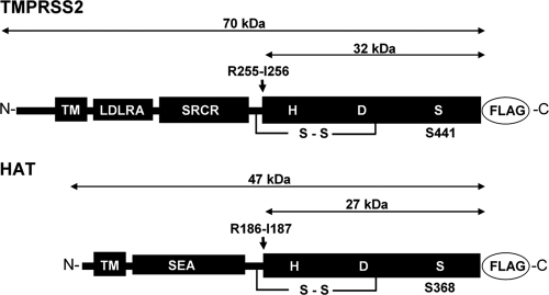
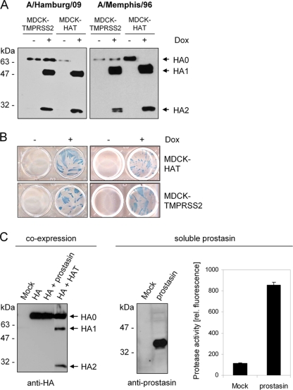
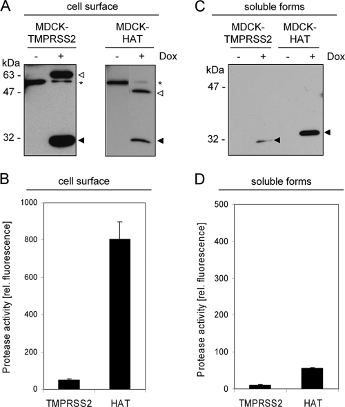
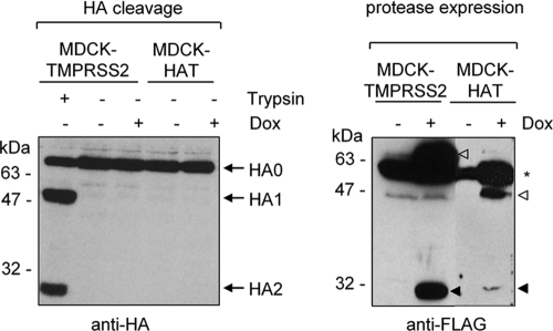
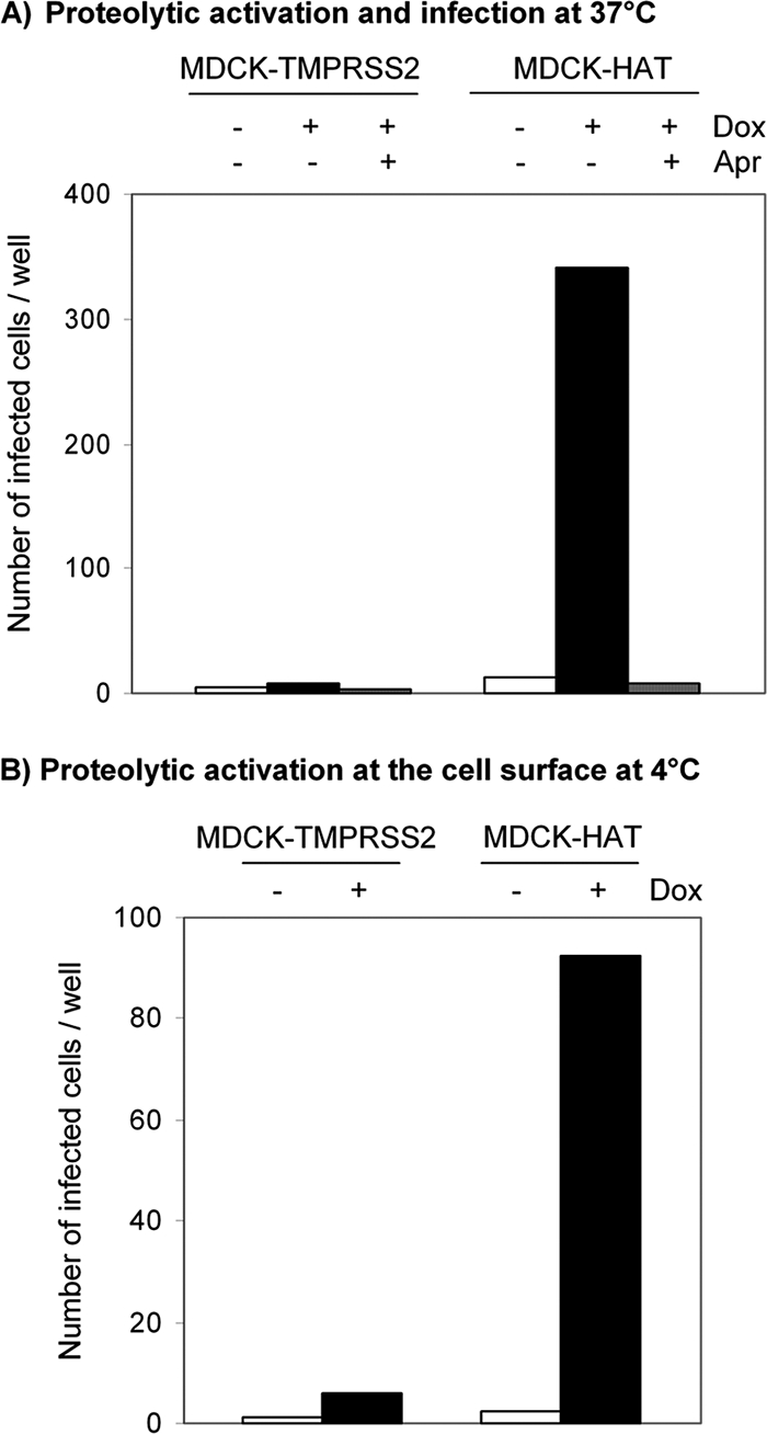
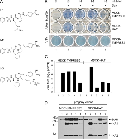
Proteolytic cleavage of the influenza virus surface glycoprotein hemagglutinin (HA) by host cell proteases is crucial for infectivity and virus spread. The proteases HAT (human airway trypsin-like protease) and TMPRSS2 (transmembrane protease serine S1 member 2) known to be present in the human airways were previously identified as proteases that cleave HA. We studied subcellular localization of HA cleavage and cleavage inhibition of seasonal influenza virus A/Memphis/14/96 (H1N1) and pandemic virus A/Hamburg/5/2009 (H1N1) in MDCK cells that express HAT and TMPRSS2 under doxycycline-induced transcriptional activation. We made the following observations: (i) HA is cleaved by membrane-bound TMPRSS2 and HAT and not by soluble forms released into the supernatant; (ii) HAT cleaves newly synthesized HA before or during the release of progeny virions and HA of incoming viruses prior to endocytosis at the cell surface, whereas TMPRSS2 cleaves newly synthesized HA within the cell and is not able to support the proteolytic activation of HA of incoming virions; and (iii) cleavage activation of HA and virus spread in TMPRSS2- and HAT-expressing cells can be suppressed by peptide mimetic protease inhibitors. The further development of these inhibitors could lead to new drugs for influenza treatment.


Figures

FIG. 1.
FIG. 1.
Schematic domain structures of HAT and TMPRSS2. HAT and TMPRSS2 are synthesized as single-chain zymogens that consist of an N-terminal transmembrane domain (TM), a stem region containing, e.g., sea urchin sperm protein, enterokinase, and agrin domain (SEA) for HAT or low-density lipoprotein receptor class A domain (LDLRA), and scavenger receptor cysteine-rich domain (SRCR) for TMPRSS2, and a C-terminal trypsin-like serine protease domain that contains the catalytic triad consisting of histidine (H), aspartic acid (D), and serine (S). The zymogens are activated by proteolytic cleavage at arginine residues R186 for HAT and R255 for TMPRSS2 (indicated by arrows). For immunochemical detection of the recombinant proteases HAT and TMPRSS2 are expressed with a C-terminally fused FLAG tag peptide (DYKDDDDK).
FIG. 2.
FIG. 2.
Proteolytic processing of HA by the proteases HAT, TMPRSS2, and prostasin. (A) MDCK-TMPRSS2 and MDCK-HAT cells were infected with H1N1 influenza viruses A/Hamburg/09 or A/Memphis/96 at an MOI of 0.01 and incubated in the absence (−) or presence (+) of doxycycline (Dox) for 24 h. Virus-containing cell supernatants were concentrated by ultracentrifugation, subjected to SDS-PAGE under reducing conditions, and subsequently transferred to a PVDF membrane and analyzed by using HA-specific antibodies and HRP-conjugated secondary antibodies. (B) MDCK-HAT and MDCK-TMPRSS2 cells were infected with A/Memphis/96 or A/Hamburg/09 at an MOI of 0.01 and incubated in the absence or presence of Dox for 24 h to allow multiple cycles of viral replication. Infected cells were immunostained against the viral nucleoprotein. (C) MDCK cells were cotransfected with pCAGGS-HA and either pCAGGS, pCAGGS-prostasin, or pCAGGS-HAT. Mock transfections were done with empty pCAGGS. At 24 h posttransfection, cell lysates were analyzed by SDS-PAGE and Western blotting with antibodies against H3 (left panel). Supernatants of pCAGGS-prostasin or pCAGGS (Mock) transfected MDCK cells were concentrated ∼100-fold and subjected to SDS-PAGE and Western blot analysis with prostasin-specific antibodies (middle panel). Enzymatic activity of the supernatants was examined by incubation with the fluorogenic substrate peptide Boc-Leu-Gly-Arg-AMC and release of AMC due to hydrolysis of the substrate was measured. The results are presented as mean enzymatic activities for three independent experiments.
FIG. 3.
FIG. 3.
Cell surface expression of TMPRSS2 and HAT, shedding from the cell surface, and determination of the enzymatic activity of membrane-bound and soluble forms. (A) MDCK-TMPRSS2 and MDCK-HAT cells were grown in 6-well plates in the absence or presence of doxycycline (Dox) for 24 h. Cell surface proteins were biotinylated; the cells were then lysed, and biotin-labeled proteins were precipitated and subjected to SDS-PAGE under reducing conditions and Western blot analysis with FLAG-specific antibodies. The zymogen and the mature form of either protease are indicated by open and filled arrowheads, respectively. A nonspecific band recognized by the FLAG-specific antiserum is indicated by an asterisk. (B) MDCK-TMPRSS2 and MDCK-HAT cells were grown in 96-well plates with or without Dox for 24 h. Protease activity at the cell surface was determined by incubation with the fluorogenic peptide substrate Boc-Leu-Gly-Arg-AMC, and the fluorescence intensity of the AMC was measured. The results are the mean enzymatic activities for three independent experiments, with values for control cells (−Dox) subtracted from values of Dox-treated cells within each experiment. (C) The soluble forms of HAT and TMPRSS2 released from MDCK-HAT and MDCK-TMPRSS2 cells grown in the absence (−) or presence (+) of Dox were concentrated and analyzed by Western blotting with FLAG-specific antibodies. (D) Enzymatic activity of concentrated TMPRSS2- or HAT-containing supernatants was measured by using the fluorogenic peptide Boc-Leu-Gly-Arg-AMC as described above.
FIG. 4.
FIG. 4.
Examination of HA cleavage by soluble HAT and TMPRSS2. MDCK-TMPRSS2 or MDCK-HAT cells grown on coverslips with or without addition of doxycycline (Dox) to the growth medium were cocultivated with transiently HA-expressing MDCK cells grown on separate coverslips next to each other in one cell culture dish for 30 h as described in Materials and Methods. As a positive control, 1 μg of trypsin/ml was added during cocultivation. Cells of each coverslip were harvested and subjected to SDS-PAGE and Western blot analysis with H3-specific antibodies (left panel) or FLAG-specific antibodies (right panel). Zymogens (open arrowheads) and the catalytic domains (filled arrowhead) of the mature forms of TMPRSS2 and HAT are indicated. A nonspecific cross-reacting band is indicated by an asterisk.
FIG. 5.
FIG. 5.
Proteolytic activation of influenza virions at the stage of entry. (A) Proteolytic activation at 37°C. MDCK-TMPRSS2 and MDCK-HAT cells grown in 96-well plates with or without doxycycline (Dox) were inoculated with human influenza virus A/Memphis/96 (H1N1) containing uncleaved HA0 (∼103 virions per well) and incubated for 10 h at 37°C to allow single cycle replication. As a control inoculation with virions was performed in the presence of 25 μM aprotinin (Apr). Cells were immunostained against NP, and the number of infected cells per well was determined. The results are the mean values of a representative experiment. (B) Proteolytic activation at 4°C prior to endocytosis. Influenza virus A/Memphis/96 containing HA0 was adsorbed to MDCK-TMPRSS2 and MDCK-HAT cells for 6 h at 4°C. Virions were removed from the cells and used for inoculation of wild-type MDCK cells seeded in 96-well plates. At 10 h p.i. MDCK cells were immunostained against NP, and infected cells per well were counted. The results are the mean values of a representative experiment.
FIG. 6.
FIG. 6.
Suppression of HA cleavage and influenza virus spread by protease inhibitors. (A) Structural formulas of peptide mimetic inhibitors I-1, I-2, and I-3. (B) MDCK-TMPRSS2 and MDCK-HAT cells were infected with A/Hamburg/09 (H1N1) at an MOI of 0.02 and VSV at an MOI of 0.001, respectively, and incubated with or without doxycycline (Dox) in the absence (Ø) or presence of inhibitor I-1, I-2, or I-3 at a final concentration of 30 μM at 37°C. At 24 h p.i. the cells were immunostained with influenza virus NP- and VSV-specific antibodies, respectively. (C) MDCK-TMPRSS2 and MDCK-HAT cells were infected with A/Hamburg/09 at an MOI of 0.01 and incubated in the absence or presence of doxycycline for 24 h (lanes 1 and 2, see panel B) in the absence or presence of inhibitor I-1 (lane 3), I-2 (lane 4), or I-3 (lane 5). Virus titers were determined by plaque assay at 24 h p.i. Note that the bars representing virus titers in cells without induction of TMPRSS2 and HAT expression (−Dox), respectively, cannot be seen in the figure since the titers were below the limit of determination (102 PFU/ml). The results are the mean values of two independent experiments. (D) Virus-containing cell supernatants shown in Fig. 6C were concentrated by ultracentrifugation and then subjected to SDS-PAGE under reducing conditions and Western blot analysis with HA-specific antibodies.

Similar articles

Cited by

References

    1. Afar, D. E., I. Vivanco, R. S. Hubert, J. Kuo, E. Chen, D. C. Saffran, A. B. Raitano, and A. Jakobovits. 2001. Catalytic cleavage of the androgen-regulated TMPRSS2 protease results in its secretion by prostate and prostate cancer epithelia. Cancer Res. 61:1686-1692. - PubMed
    1. Becker, G. L., F. Sielaff, M. E. Than, I. Lindberg, S. Routhier, R. Day, Y. Lu, W. Garten, and T. Steinmetzer. 2010. Potent inhibitors of furin and furin-like proprotein convertases containing decarboxylated P1 arginine mimetics. J. Med. Chem. 53:1067-1075. - PMC - PubMed
    1. Böttcher, E., C. Freuer, T. Steinmetzer, H. D. Klenk, and W. Garten. 2009. MDCK cells that express proteases TMPRSS2 and HAT provide a cell system to propagate influenza viruses in the absence of trypsin and to study cleavage of HA and its inhibition. Vaccine 27:6324-6329. - PubMed
    1. Böttcher, E., T. Matrosovich, M. Beyerle, H. D. Klenk, W. Garten, and M. Matrosovich. 2006. Proteolytic activation of influenza viruses by serine proteases TMPRSS2 and HAT from human airway epithelium. J. Virol. 80:9896-9898. - PMC - PubMed
    1. Boycott, R., H. D. Klenk, and M. Ohuchi. 1994. Cell tropism of influenza virus mediated by hemagglutinin activation at the stage of virus entry. Virology 203:313-319. - PubMed

Publication types

MeSH terms

LinkOut - more resources