Phages harboring specific peptides that recognize the N protein of the porcine reproductive and respiratory syndrome virus distinguish the virus from other viruses
- PMID: 20237096
- PMCID: PMC2863871
- DOI: 10.1128/JCM.01707-09
Phages harboring specific peptides that recognize the N protein of the porcine reproductive and respiratory syndrome virus distinguish the virus from other viruses
Abstract
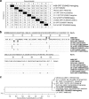
The aim of the current study was to develop a novel diagnostic test for detecting porcine reproductive and respiratory syndrome virus (PRRSV) using phage display technology. The N gene of PRRSV isolate HH08 was cloned following reverse transcription-PCR. Sequence comparison indicated that the N gene shared 96.4% homology to that of North American PRRSV (isolate VR2332) and 35.5% with that of European PRRSV (isolate LV), indicating that the PRRSV isolate was related to the North American PRRSV genotype. The bacterially expressed N protein was used as a target in a biopanning process using a phage display random peptide library. Seven phages expressing different peptides had a specific binding activity with the N protein. The putative binding motifs were identified by DNA sequencing. More importantly, the selected phages harboring specific peptides that recognize the N protein of PRRSV were able to efficiently distinguish PRRSV from other viruses in enzyme-linked immunosorbent assays.
Figures


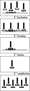
 ” symbol indicates the specific peptides on the ends of the phages; “
 ” indicates target proteins; “▧” indicates immobilized support for a target. Other symbols on the ends of phages indicate unspecific exogenous peptides.


Similar articles
-
[Screening of polypeptides binding to porcine reproductive and respiratory syndrome virus by phage display library].Wei Sheng Wu Xue Bao. 2011 Jan;51(1):127-33. Wei Sheng Wu Xue Bao. 2011. PMID: 21465799 Chinese.
-
High risk of false positive results in a widely used diagnostic test for detection of the porcine reproductive and respiratory syndrome virus (PRRSV).Vet Microbiol. 2006 Jun 15;115(1-3):21-31. doi: 10.1016/j.vetmic.2006.01.001. Epub 2006 Feb 3. Vet Microbiol. 2006. PMID: 16458457
-
Performance of ELISA antigens prepared from 8 isolates of porcine reproductive and respiratory syndrome virus with homologous and heterologous antisera.Can J Vet Res. 1997 Oct;61(4):299-304. Can J Vet Res. 1997. PMID: 9342455 Free PMC article.
-
Epitope specificity of monoclonal antibodies to the N-protein of porcine reproductive and respiratory syndrome virus determined by ELISA with synthetic peptides.Vet Immunol Immunopathol. 2005 Mar 10;104(1-2):59-68. doi: 10.1016/j.vetimm.2004.10.004. Vet Immunol Immunopathol. 2005. PMID: 15661331
-
Porcine reproductive and respiratory syndrome virus (PRRSV) in serum and oral fluid samples from individual boars: will oral fluid replace serum for PRRSV surveillance?Virus Res. 2010 Dec;154(1-2):170-6. doi: 10.1016/j.virusres.2010.07.025. Epub 2010 Jul 27. Virus Res. 2010. PMID: 20670665 Review.
Cited by
-
Transmissible gastroenteritis virus: identification of M protein-binding peptide ligands with antiviral and diagnostic potential.Antiviral Res. 2013 Sep;99(3):383-90. doi: 10.1016/j.antiviral.2013.06.015. Epub 2013 Jul 2. Antiviral Res. 2013. PMID: 23830854 Free PMC article.
-
Phage display for identifying peptides that bind the spike protein of transmissible gastroenteritis virus and possess diagnostic potential.Virus Genes. 2015 Aug;51(1):51-6. doi: 10.1007/s11262-015-1208-7. Epub 2015 May 27. Virus Genes. 2015. PMID: 26013256 Free PMC article.
-
Expression of the nucleocapsid protein of porcine reproductive and respiratory syndrome virus in soybean seed yields an immunogenic antigenic protein.Planta. 2012 Mar;235(3):513-22. doi: 10.1007/s00425-011-1523-8. Epub 2011 Oct 5. Planta. 2012. PMID: 21971995
-
Development of a porcine epidemic diarrhea virus M protein-based ELISA for virus detection.Biotechnol Lett. 2011 Feb;33(2):215-20. doi: 10.1007/s10529-010-0420-8. Epub 2010 Sep 30. Biotechnol Lett. 2011. PMID: 20882317 Free PMC article.
-
Phage displayed peptides to avian H5N1 virus distinguished the virus from other viruses.PLoS One. 2011;6(8):e23058. doi: 10.1371/journal.pone.0023058. Epub 2011 Aug 22. PLoS One. 2011. PMID: 21887228 Free PMC article.
References
-
- An, T. Q., Y. J. Zhou, G. Q. Liu, Z. J. Tian, J. Li, H. J. Qiu, and G. Z. Tong. 2007. Genetic diversity and phylogenetic analysis of glycoprotein 5 of PRRSV isolates in mainland China from 1996 to 2006: coexistence of two NA-subgenotypes with great diversity. Vet. Microbiol. 123:43-52. - PubMed
-
- Cavanagh, D. 1997. Nidovirales: a new order comprising Coronaviridae and Arteriviridae. Arch. Virol. 142:629-633. - PubMed
-
- Chames, P., and D. Baty. 2000. Antibody engineering and its applications in tumor targeting and intracellular immunization. FEMS Microbiol. Lett. 189:1-8. - PubMed
Publication types
MeSH terms
Substances
Associated data
- Actions
LinkOut - more resources
Full Text Sources

