

Case Reports
doi: 10.1128/JCM.02518-09.
Epub 2010 Mar 17.
Tuberculosis in alpacas (Lama pacos) caused by Mycobacterium bovis
Affiliations
- PMID: 20237097
- PMCID: PMC2863934
- DOI: 10.1128/JCM.02518-09
Item in Clipboard
Case Reports
Tuberculosis in alpacas (Lama pacos) caused by Mycobacterium bovis
J Clin Microbiol.
2010 May.
Abstract
We report three cases of tuberculosis in alpacas from Spain caused by Mycobacterium bovis. The animals revealed two different lesional patterns. Mycobacterial culture and PCR assay yielded positive results for M. bovis. Molecular typing of the isolates identified spoligotype SB0295 and identical variable-number tandem repeat (VNTR) allele sizes.
Figures
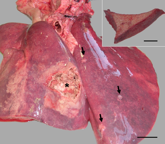
Multifocal areas of granulomatous, caseous bronchopneumonia (arrows) with a large area of bronchopneumonia communicating with the bronchial lumen (asterisk). Bar = 5 cm. The inset image shows a diffuse pattern of granulomatous pneumonia observed in alpaca 1. Bar = 4.5 cm.
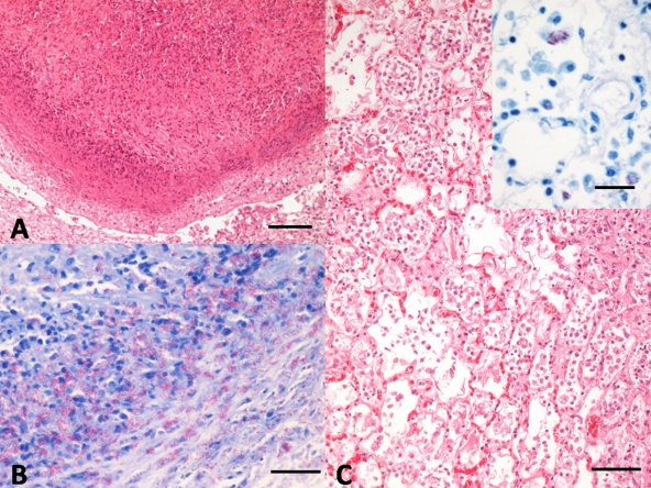
(A) Alpaca 1. Nodular granulomatous lesion composed of a central core of necrosis, surrounded by degenerated neutrophils and cell debris, epithelioid macrophages, and scattered lymphocytes and plasma cells. Note the manifest capsule of connective tissue delimiting the lesion (as shown by hematoxylin and eosin [H&E] staining). Bar = 100 μm. (B) Alpaca 1. Abundant acid-fast bacteria in the periphery of the granulomatous lesion. Ziehl-Neelsen staining. Bar = 20 μm. (C) Alpaca 3. Marked proliferation of histiocytes, with diffuse intraalveolar infiltrate of foamy macrophages and epithelioid cells. H-E. Bar = 50 μm. The inset image shows random foamy macrophages, laden with numerous acid-fast bacteria within their cytoplasm. Ziehl-Neelsen staining. Bar = 20 μm.
References
-
- Barlow, A. M., K. A. Mitchell, and K. H. Visram. 1999. Bovine tuberculosis in llama (Lama glama) in the UK. Vet. Rec. 145:639-640. - PubMed
-
- Casweel, J. L., and K. J. Williams. 2007. Respiratory system, p. 606-610. In M. G. Maxie (ed.), Jubb, Kennedy, and Palmer's pathology of domestic animals, vol. 2, 5th ed. Elsevier-Saunders, Philadelphia, PA.
-
- Cousins, D. V., and N. Florisson. 2005. A review of tests available for use in the diagnosis of tuberculosis in non-bovine species. Rev. Sci. Tech. 24:1039-1059. - PubMed
-
- Cousins, D. V. 2001. Mycobacterium bovis infection and control in domestic livestock. Rev. Sci. Tech. 20:71-85. - PubMed
Publication types
MeSH terms
LinkOut - more resources
Full Text Sources
Medical
