. 2010 May 1;21(9):1519-29.
doi: 10.1091/mbc.e10-01-0008. Epub 2010 Mar 17.

A regulated complex of the scaffolding proteins PDZK1 and EBP50 with ezrin contribute to microvillar organization

Affiliations

A regulated complex of the scaffolding proteins PDZK1 and EBP50 with ezrin contribute to microvillar organization

David P LaLonde et al. Mol Biol Cell. .

Abstract

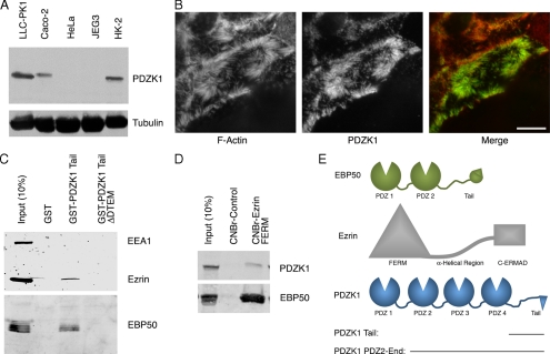
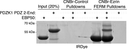
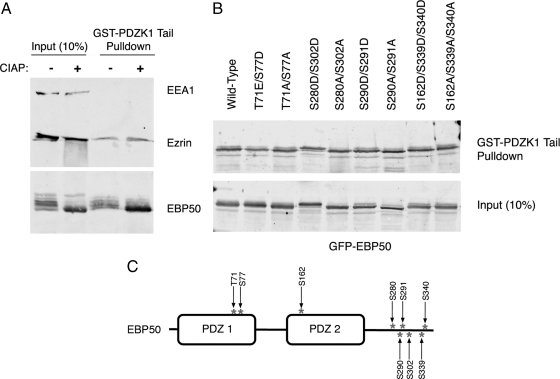
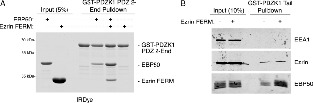
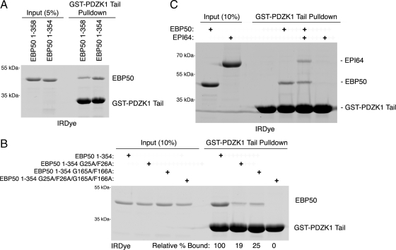
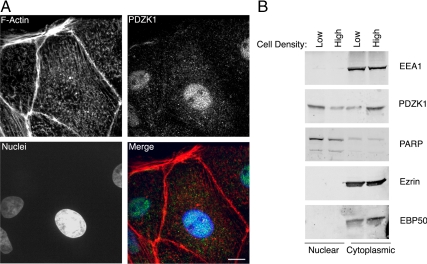
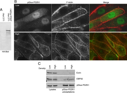
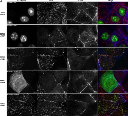
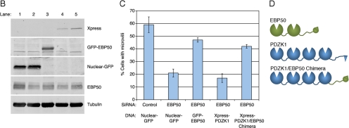
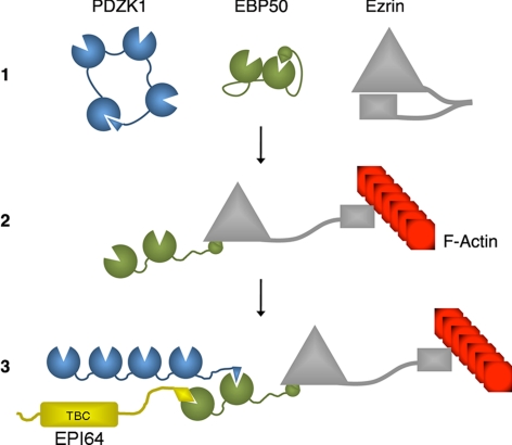
PDZK1 and ezrin, radixin, moesin binding phosphoprotein 50 kDa (EBP50) are postsynaptic density 95/disc-large/zona occludens (PDZ)-domain-containing scaffolding proteins found in the apical microvilli of polarized epithelial cells. Binary interactions have been shown between the tail of PDZK1 and the PDZ domains of EBP50, as well as between EBP50 and the membrane-cytoskeletal linking protein ezrin. Here, we show that these molecules form a regulated ternary complex in vitro and in vivo. Complex formation is cooperative because ezrin positively influences the PDZK1/EBP50 interaction. Moreover, the interaction of PDZK1 with EBP50 is enhanced by the occupancy of EBP50's adjacent PDZ domain. The complex is further regulated by location, because PDZK1 shuttles from the nucleus in low confluence cells to microvilli in high confluence cells, and this regulates the formation of the PDZK1/EBP50/ezrin complex in vivo. Knockdown of EBP50 decreases the presence of microvilli, a phenotype that can be rescued by EBP50 re-expression or expression of a PDZK1 chimera that is directly targeted to ezrin. Thus, when appropriately located, PDZK1 can provide a function necessary for microvilli formation normally provided by EBP50. By entering into the ternary complex, PDZK1 can both enhance the scaffolding at the apical membrane as well as augment EBP50's role in microvilli formation.


Figures

Figure 1.
Figure 1.
PDZK1 is found in microvilli and forms a complex with EBP50 and ezrin. (A) Immunoblot with PDZK1 antibody of lysates from the indicated cell lines, with tubulin used as a loading control. (B) Immunofluorescence localization of PDZK1 to microvilli in LLC-PK1 cells. F-actin was labeled with rhodamine phalloidin. The image represents a maximum projection of the apical aspect of the cell. Bar, 10 μm. (C) The GST constructs indicated were used for precipitations from JEG3 cell lysates. Retained and eluted material was blotted for ezrin and EBP50, with EEA1 serving as a negative control. (D) The ezrin FERM domain coupled to Sepharose or control Sepharose beads were used to precipitate proteins from LLC-PK1 cells. Both EBP50 and PDZK1 were coprecipitated with the FERM domain. (E) Schematics of PDZK1, EBP50 and ezrin are shown with the relevant domains and truncation constructs indicated.
Figure 2.
Figure 2.
EBP50 serves as a linker between PDZK1 and ezrin. In vitro binding assays were performed between the indicated CNBr resin-bound proteins and soluble proteins. Although the FERM domain strongly precipitates EBP50, it only precipitates PDZK1 PDZ 2-End when EBP50 is also present. These results indicate the existence of a PDZK1–EBP50–ezrin ternary complex with EBP50 as the intermediate component.
Figure 3.
Figure 3.
EBP50 phosphorylation does not significantly impact its association with PDZK1. (A) JEG3 cell extracts were treated with or without CIAP then subjected to pull-downs with the resin-bound GST-PDZK1 tail. Dephosphorylation of EBP50, as indicated by the loss of bands of different mobility by SDS-PAGE, did not alter the association with the GST-PDZK1 tail. (B) The indicated phosphomimetic and phosphodeficient EBP50 mutants were expressed in JEG3 cells. Lysates were prepared and subjected to pull-downs with the resin-bound GST-PDZK1 tail. All constructs bound the PDZK1 tail comparably. (C) Schematic of EBP50 showing the location of phosphorylation sites that were mutated.
Figure 4.
Figure 4.
The ezrin FERM domain positively regulates the association between EBP50 and PDZK1. (A) In vitro binding assays were performed between resin-bound GST-PDZK1 PDZ 2-End versus soluble EBP50 or ezrin FERM in the indicated combinations. The FERM domain both coprecipitates with PDZK1 when EBP50 is present and increases the affinity of EBP50 for PDZK1. (B) Resin-bound GST-PDZK1 tail was used to precipitate proteins from JEG3 cell extracts with either BSA or soluble ezrin FERM domain added to the lysates. EBP50 binds the GST-PDZK1 tail more efficiently in the presence of excess ezrin FERM domain.
Figure 5.
Figure 5.
Characterization of the interaction between PDZK1 and the EBP50 PDZ domains. (A) In vitro binding assays were performed between resin-bound GST-PDZK1 tail and the indicated EBP50 constructs. EBP50 lacking its final four residues (1-354) binds PDZK1 more efficiently than full-length EBP50 (1–358). (B) In vitro binding assays were performed between resin-bound GST-PDZK1 tail and soluble EBP50 with mutated, inactive PDZ domains (G25A/F26A has an inactive PDZ1, whereas G165A/F166A has an inactive PDZ2). The relative amounts of PDZ mutants bound compared with the nonmutated EBP50 construct are shown below the gel. Mutation of either PDZ domain reduces the association between PDZK1 and EBP50. (C) In vitro binding assays were performed between resin-bound GST-PDZK1 tail and the indicated constructs. EPI64, a strong binder of EBP50 PDZ1, is able to precipitate with the GST-PDZK1 tail in the presence of EBP50. The results of B and C indicate that PDZK1 can bind PDZ2 of EBP50 more efficiently when PDZ1 is occupied.
Figure 6.
Figure 6.
PDZK1 cycles into and out of the nucleus based on cell confluence. (A) Immunofluorescence localization of endogenous PDZK1 in the nucleus in low confluence LLC-PK1 cells costained to show F-actin and cell nuclei. The image represents a maximum projection through the cell. Bar, 10 μm. (B) LLC-PK1 cells at either high or low confluence were fractionated into nuclear and cytoplasmic fractions then blotted for the indicated proteins. PDZK1 becomes less enriched in the nucleus in high confluence cells, whereas EBP50 and ezrin are not found in the nuclear fraction.
Figure 7.
Figure 7.
Cell confluence regulates the association between PDZK1 and ezrin/EBP50. (A) Lysates from cells stably expressing pGlue-PDZK1 and parental cells were blotted with monoclonal anti-HA antibody to show expression of the pGlue construct. (B) Immunofluorescence localization of stably expressed pGlue-tagged PDZK1 in LLC-PK1 cells at high and low confluence costained with rhodamine phalloidin to show F-actin. The tagged version of PDZK1 exhibits similar nuclear shuttling to the endogenous protein. (C) pGlue-PDZK1 was precipitated out of the stably expressing cell line at both high and low confluence and analyzed by SDS-PAGE. Twice as much eluate was analyzed for the ezrin and EBP50 blots as was used for the HA blot for PDZK1. EBP50 and ezrin are both found to coprecipitate with PDZK1 more efficiently under higher cell confluence.
Figure 8.
Figure 8.
PDZK1 exhibits functional redundancy with EBP50 in microvilli formation. (A) JEG3 cells were transfected with the indicated constructs, treated with siRNA to EBP50 or a control luciferase oligonucleotide one day later, allowed to grow for 2 d and then processed for immunofluorescence. Bar, 10 μm. (B) Lysates of the conditions listed in A and C were blotted as indicated. The lane numbers correspond to the five conditions represented in A moving from top to bottom or on the graph in C moving from left to right. (C) Percentage of cells exhibiting microvilli under the indicated conditions was quantified. EBP50 knockdown inhibits microvilli formation and this is significantly (p < 0.01) rescued by both EBP50 and a PDZK1/EBP50 chimera but not by wild-type PDZK1. (D) Schematic of the domains present in the PDZK1/EBP50 chimera versus the wild-type proteins.
Figure 8.
Figure 8.
PDZK1 exhibits functional redundancy with EBP50 in microvilli formation. (A) JEG3 cells were transfected with the indicated constructs, treated with siRNA to EBP50 or a control luciferase oligonucleotide one day later, allowed to grow for 2 d and then processed for immunofluorescence. Bar, 10 μm. (B) Lysates of the conditions listed in A and C were blotted as indicated. The lane numbers correspond to the five conditions represented in A moving from top to bottom or on the graph in C moving from left to right. (C) Percentage of cells exhibiting microvilli under the indicated conditions was quantified. EBP50 knockdown inhibits microvilli formation and this is significantly (p < 0.01) rescued by both EBP50 and a PDZK1/EBP50 chimera but not by wild-type PDZK1. (D) Schematic of the domains present in the PDZK1/EBP50 chimera versus the wild-type proteins.
Figure 9.
Figure 9.
Schematic of the PDZK1–EBP50–ezrin complex. 1. The microvillar scaffold proteins PDZK1, EBP50, and ezrin exist in intramolecularly associated conformations before entering into a complex. These conformations reduce their associations with one another. 2. Ezrin becomes “activated” or “open” through a combination of membrane binding and phosphorylation. This allows its amino-terminal FERM domain to interact with EBP50 and its carboxy-terminal C-ERMAD to interact with F-actin. 3. The association with ezrin locks the EBP50 in an open conformation, thereby enhancing the association between EBP50 and the tail of PDZK1. PDZK1, EBP50 and ezrin are then able to form a complex. Notably, the tail of PDZK1 must be released from its intramolecular interaction with its own first PDZ domain to interact with EBP50. Additional EBP50 binding partners, such as EPI64 (labeled with its amino-terminal TBC domain), may also enter into this macromolecular complex.

Similar articles

Cited by

References

    1. Angers S., Thorpe C. J., Biechele T. L., Goldenberg S. J., Zheng N., MacCoss M. J., Moon R. T. The KLHL12-Cullin-3 ubiquitin ligase negatively regulates the Wnt-beta-catenin pathway by targeting Dishevelled for degradation. Nat. Cell Biol. 2006;8:348–357. - PubMed
    1. Bretscher A., Edwards K., Fehon R. G. ERM proteins and merlin: integrators at the cell cortex. Nat. Rev. Mol. Cell Biol. 2002;3:586–599. - PubMed
    1. Cao T. T., Deacon H. W., Reczek D., Bretscher A., von Zastrow M. A kinase-regulated PDZ-domain interaction controls endocytic sorting of the beta2-adrenergic receptor. Nature. 1999;401:286–290. - PubMed
    1. Donowitz M., Cha B., Zachos N. C., Brett C. L., Sharma A., Tse C. M., Li X. NHERF family and NHE3 regulation. J. Physiol. 2005;567:3–11. - PMC - PubMed
    1. Doyle D. A., Lee A., Lewis J., Kim E., Sheng M., MacKinnon R. Crystal structures of a complexed and peptide-free membrane protein-binding domain: molecular basis of peptide recognition by PDZ. Cell. 1996;85:1067–1076. - PubMed

Publication types

MeSH terms