. 2010 May 1;21(9):1556-68.
doi: 10.1091/mbc.e09-12-1002. Epub 2010 Mar 17.

Yip1A structures the mammalian endoplasmic reticulum

Affiliations

Yip1A structures the mammalian endoplasmic reticulum

Kaitlyn M Dykstra et al. Mol Biol Cell. .

Abstract

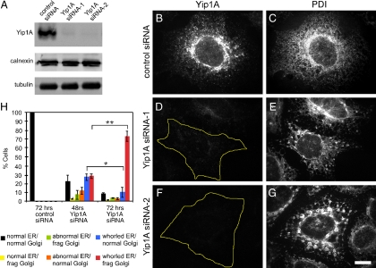
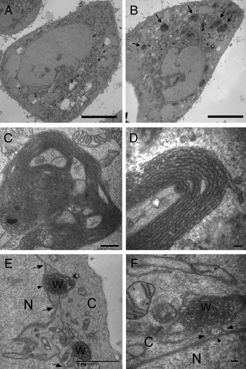
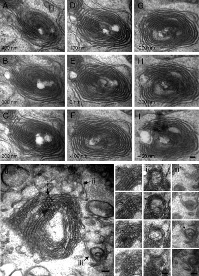
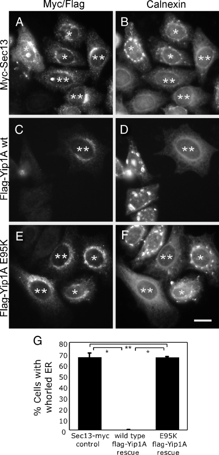
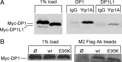
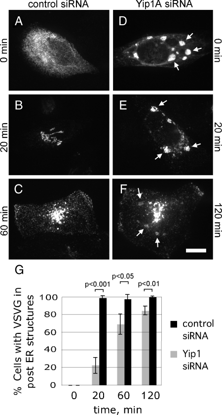
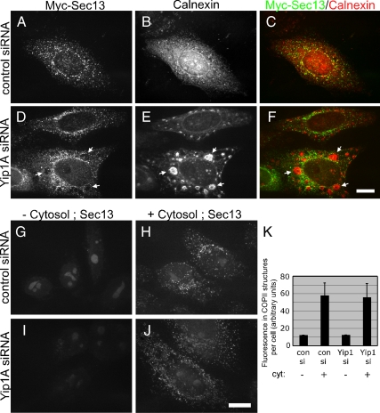
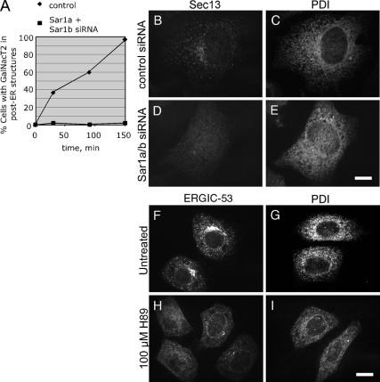
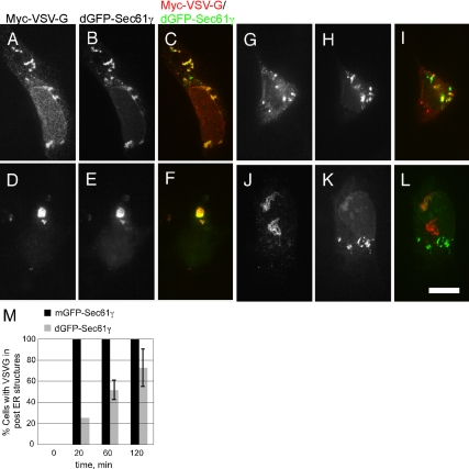
The structure of the endoplasmic reticulum (ER) undergoes highly regulated changes in specialized cell types. One frequently observed type of change is its reorganization into stacked and concentrically whorled membranes, but the underlying mechanisms and functional relevance for cargo export are unknown. Here, we identify Yip1A, a conserved membrane protein that cycles between the ER and early Golgi, as a key mediator of ER organization. Yip1A depletion led to restructuring of the network into multiple, micrometer-sized concentric whorls. Membrane stacking and whorl formation coincided with a marked slowing of coat protein (COP)II-mediated protein export. Furthermore, whorl formation driven by exogenous expression of an ER protein with no role in COPII function also delayed cargo export. Thus, the slowing of protein export induced by Yip1A depletion may be attributed to a proximal role for Yip1A in regulating ER network dispersal. The ER network dispersal function of Yip1A was blocked by alteration of a single conserved amino acid (E95K) in its N-terminal cytoplasmic domain. These results reveal a conserved Yip1A-mediated mechanism for ER membrane organization that may serve to regulate cargo exit from the organelle.


Figures

Figure 1.
Figure 1.
RNAi-mediated Yip1A depletion causes ER compaction. (A) Immunoblot demonstrating loss of Yip1A by two distinct siRNAs. HeLa cells stably expressing Golgi-localized GFP-GalNacT2 were transfected with a control siRNA, Yip1A siRNA-1, or Yip1A siRNA-2; harvested 72 h later; and probed using antibodies against Yip1A, calnexin, and tubulin. (B–G) Yip1A loss correlates with ER morphological changes. Cells transfected with control siRNA (B and C), Yip1A siRNA-1 (D and E), or Yip1A siRNA-2 (F and G) were fixed 72 h later and doubly stained with antibodies against Yip1A (B, D, and F) and PDI (C, E, and G). Bar, 10 μm. (H) ER morphological changes precede Golgi fragmentation. Cells transfected with a control siRNA or Yip1 siRNA-2 were fixed 48 or 72 h later and stained with antibodies against PDI. ER and Golgi morphologies were classified as indicated (frag Golgi, fragmented Golgi; whorled ER indicates compacted ER as shown in E and G; abnormal ER indicates ER morphologies intermediate between normal and whorled). Quantitation of the average percentage of cells displaying the indicated ER and Golgi morphologies from three independent experiments (>50 cells/condition/experiment) is shown, ±SD. Single asterisk indicates p < 0.05 and double asterisk indicates p < 0.001 (Student's t test).
Figure 2.
Figure 2.
Ultrastructural analysis of cells lacking Yip1A. (A and B) A low-magnification thin section transmission EM view of cells treated with a control (A) or Yip1A (B) siRNA. Arrows (B) indicate dense ER membrane aggregates seen only in Yip1A siRNA-treated cells. Bar, 10 μm. (C) A higher magnification view of an entire ER whorl. Bar, 500 nm. (D) A high magnification view of stacked membranes of a portion of an ER whorl. Bar, 100 nm. (E and F) ER whorls are continuous with the nuclear envelope. A low-magnification view of two interconnected whorls each connected to the nuclear envelope (E) and a higher magnification view of a whorl exhibiting connections to the outer nuclear envelope (F). Arrowheads (E and F) indicate the nuclear envelope and arrows (E and F) indicate membrane continuities between whorl and nuclear envelope. N, nucleus; C, cytoplasm; W, whorl. Bar, 2 μm (E) and 100 nm (F).
Figure 3.
Figure 3.
Early ER whorls consist of both sheets and tubules. (A–I) Serial (100-nm) transmission EM thin sections through an ER whorl 48 h after Yip1A siRNA treatment. Bar, 100 nm. (J) A thin section micrograph through a different ER whorl 48 h after Yip1A siRNA treatment. Arrows (i, ii, and iii) indicate three different examples of tubular morphology. Serial 100-nm thin sections through the corresponding regions (i, ii, and iii) of the whorl in J are shown in i', ii', and iii'.
Figure 4.
Figure 4.
The whorled ER phenotype is rescued by a wild type but not mutant siRNA-immune Yip1A construct. Cells cotransfected with Yip1A siRNA-2 and either a control Sec13-Myc construct (A and B), a siRNA-immune wild-type FLAG-Yip1A construct (C and D), or a siRNA-immune mutant (E95K) FLAG-Yip1A construct (E and F) were fixed 72 h later and doubly stained with antibodies against the Myc epitope (A) and calnexin (B) or the FLAG epitope (C and E) and calnexin (D and F). Single asterisks (A–F) indicate expressing cells that exhibit ER whorls. Double asterisks (A–F) indicate expressing cells that do not exhibit ER whorls. Bar, 10 μm. (G) Quantitation of the percentage of Sec13-Myc or wild type or mutant (E95K) FLAG-Yip1A–expressing cells displaying the whorled ER phenotype from three independent experiments (>50 cells per experiment), ±SD. Single asterisks indicate p < 0.0001 (Student's t test). Double asterisk indicates no statistically significant difference.
Figure 5.
Figure 5.
Both wild-type and E95K Yip1A bind to DP1. (A) HeLa cells transfected with either Myc-DP1 or Myc-DP1L1 were solubilized in 1% Triton X-100 and subjected to immunoprecipitation with either a control antibody (IgG) or Yip1A antibody. Bound protein was subjected to immunoblotting with an antibody against the Myc epitope. (B) HeLa cells cotransfected with Myc-DP1 and either wild-type FLAG-Yip1A or FLAG-Yip1A (E95K) were solubilized in 1% Triton X-100 and subjected to immunoprecipitation with M2 FLAG antibody beads. Bound protein was subjected to immunoblotting with the Myc epitope antibody. As a control, the Myc-DP1 recovered on M2 beads in the absence of FLAG-Yip1A is also shown.
Figure 6.
Figure 6.
Export of ts045 VSV-G from the ER is slowed by ER whorling. (A–F) Cells cotransfected with Myc-ts045 VSV-G and either a control siRNA (A–C) or Yip1A siRNA-2 (D–F) were shifted 48 h after transfection to 40°C to accumulate VSV-G in the ER. After an additional 24 h, cells were shifted to 32°C for 0 (A and D), 20 (B and E), 60 (C), or 120 (F) min to allow ER export. Cells were fixed and doubly stained with antibodies against the Myc epitope and calnexin (only the Myc staining is shown). Arrows (D–F) indicate the positions of ER whorls as marked by Calnexin staining. Bar, 10 μm. (G) Quantitation of the percent of cells expressing Myc-ts045 VSV-G with the protein in post-ER structures. For cells transfected with Yip1A siRNA, only cells with whorled ER, as marked by Calnexin staining, were quantified. Shown are the averages from three independent experiments, ±SD (p values obtained using the Student's t test).
Figure 7.
Figure 7.
COPII recruitment is not blocked in cells lacking Yip1A. (A–F) Cells cotransfected with Sec13-Myc and either a control siRNA (A–C) or Yip1A siRNA-2 (D–F) were fixed 72 h later and doubly stained with antibodies against the Myc epitope (A and D) and calnexin (B and E). The merge is also shown (C and F). Arrows (D–F) indicate the lack of Sec13-Myc in ER whorls. Bar, 10 μm. (G–K) Cytosol dependent COPII assembly does not require Yip1A. Cells doubly transfected with a control (G and H) or Yip1A siRNA-2 (I and J) were permeabilized with 30 μg/ml digitonin 72 h after the second transfection and incubated with an ATP-regenerating system and guanosine 5′-O-(3-thio)triphosphate in the absence (G and I) or presence (H and J) of 4 mg/ml rat liver cytosol. After 20 min at 37°C, cells were fixed and stained with antibodies against Sec13. Bar, 10 μm. (K) Quantitation of the total fluorescence in Sec13-positive structures under the indicated conditions is shown, three independent experiments, ±SD.
Figure 8.
Figure 8.
ER export blockade is not sufficient to cause ER whorling (A–E). (A) Sar1 knockdown blocks ER export. HeLa cells expressing the Golgi marker GFP-GalNacT2 and transfected with a control siRNA or siRNAs targeting both Sar1a and Sar1b isoforms were treated with 2.5 μg/ml BFA for 30 min to redistribute GFP-GalNacT2 to the ER and subsequently incubated without drug to allow ER export. At the indicated times, cells were fixed, and the percentage of cells with GFP-GalNacT2 in post-ER structures was counted. (B–E) ER export blockade by Sar1 knockdown does not affect ER morphology. Control (B and C) or Sar1 knockdown cells (D and E) were fixed 72 h after transfection and doubly stained with antibodies against Sec13 (B and D) and PDI (C and E). Bar, 10 μm. (F–I) ER export blockade by H89 treatment does not affect ER morphology. Untreated cells (F and G) or cells treated for 20 min with 100 μM H89 (H and I) were fixed and stained singly for ERGIC-53 (F and H) or PDI (G and I). Bar, 10 μm.
Figure 9.
Figure 9.
ER membrane stacking is sufficient to slow ER export. Cells cotransfected with Myc-ts045 VSV-G and either a control mGFP-Sec61γ construct (not shown but quantified in M) or the ER stack-inducing dGFP-Sec61γ construct were shifted to 40°C to accumulate VSV-G in the ER. Thereafter, cells were shifted to 32°C for 0 (A–C), 20 (D–F), 60 (G–I), or 120 (J–L) min. At the indicated times, cells were fixed and stained with Myc epitope antibodies. The corresponding VSV-G (A, D, G, and J) and GFP-Sec61γ (B, E, H, and K) as well as merged images (C, F, I, and L) are shown. Bar, 10 μm. (M) Quantitation of the kinetics of VSV-G export under each condition, the average of two independent experiments, ±SD is shown.

Similar articles

Cited by

References

    1. Ahluwalia N., Bergeron J. J., Wada I., Degen E., Williams D. B. The p88 molecular chaperone is identical to the endoplasmic reticulum membrane protein, calnexin. J. Biol. Chem. 1992;267:10914–10918. - PubMed
    1. Amarilio R., Ramachandran S., Sabanay H., Lev S. Differential regulation of endoplasmic reticulum structure through VAP-Nir protein interaction. J. Biol. Chem. 2005;280:5934–5944. - PubMed
    1. Anderson R. G., Orci L., Brown M. S., Garcia-Segura L. M., Goldstein J. L. Ultrastructural analysis of crystalloid endoplasmic reticulum in UT-1 cells and its disappearance in response to cholesterol. J. Cell Sci. 1983;63:1–20. - PubMed
    1. Aridor M., Balch W. E. Kinase signaling initiates coat complex II (COPII) recruitment and export from the mammalian endoplasmic reticulum. J. Biol. Chem. 2000;275:35673–35676. - PubMed
    1. Barr F. A., Puype M., Vandekerckhove J., Warren G. GRASP65, a protein involved in the stacking of Golgi cisternae. Cell. 1997;91:253–262. - PubMed

Publication types

Substances