. 2010 May 1;21(9):1585-96.
doi: 10.1091/mbc.e09-07-0561. Epub 2010 Mar 17.

Nup98-homeodomain fusions interact with endogenous Nup98 during interphase and localize to kinetochores and chromosome arms during mitosis

Affiliations

Nup98-homeodomain fusions interact with endogenous Nup98 during interphase and localize to kinetochores and chromosome arms during mitosis

Songli Xu et al. Mol Biol Cell. .

Abstract

Chromosomal translocations involving the Nup98 gene are implicated in leukemias, especially acute myelogenous leukemia. These translocations generate chimeric fusion proteins, all of which have in common the N-terminal half of Nup98, which contains the nucleoporin FG/GLFG repeat motifs. The homeodomain group of Nup98 fusion proteins retain the C-terminus of a homeodomain transcription factor, including the homeobox responsible for DNA binding. Current models for Nup98 leukemogenesis invoke aberrant transcription resulting from recruitment of coregulators by the Nup98 repeat domain. Here we have investigated the behavior of Nup98-homeodomain fusion proteins throughout the cell cycle. At all stages, the fusion proteins exhibit a novel localization distinct from the component proteins or fragments. During interphase, there are dynamic interactions between the Nup98 fusions and endogenous Nup98 that lead to mislocalization of the intranuclear fraction of Nup98, but do not alter the level of Nup98 at the nuclear pore complex. During mitosis, no interaction between the fusion proteins and endogenous Nup98 is observed. However, the fusions are entirely concentrated at kinetochores and on chromosome arms, sites where the APC/C, a target of Nup98 regulation, is also found. Our observations suggest new possibilities for misregulation by which Nup98 translocations may contribute to cellular transformation and leukemogenesis.


Figures

Figure 1.
Figure 1.
Nup98 leukemic fusion proteins are directed to the same intranuclear sites, distinct from the localization of the contributing proteins or domains. (A) Schematic of Nup98, the HoxA9, PMX1, and HoxD13 homeodomain proteins, and the corresponding Nup98-fusion proteins. Repeat domains containing primarily FG or GLFG repeats are indicated. GBD, binding site for Gle2/Rae1; HD, homeodomain. The site of autoproteolytic cleavage is indicated. (B) Localization of Nup98, the Nup98 repeat domain (GLFG), the HoxA9, PMX1, and HoxD13 homeodomain proteins, and the corresponding Nup98-fusion proteins. Proteins were tagged at the N-terminus with GFP, expressed in HeLa cells, and fixed for analysis with 4% paraformaldehyde. Insets in panels b–d show higher magnification of fusion protein foci. (C) Nup98-homeodomain fusions colocalize within the nucleus. CFP-tagged (false colored in red) and GFP- or YFP-tagged (false colored in green) fusion proteins were coexpressed in HeLa cells and visualized by confocal microscopy. Merged signal is shown at right. Scale bars, 5 μm.
Figure 2.
Figure 2.
The dynamic behavior of Nup98-homeodomain fusions in live cells is distinct from that of the parental proteins. (A) Left, GFP-tagged Nup98 in GLFG bodies or GFP-tagged homeodomain proteins in the nucleoplasm were photobleached and fluorescence recovery was recorded over time. Right, GFP-Nup98, the GFP-tagged Nup98 fragment corresponding to the leukemic fusion proteins (amino acids 1-469), or GFP-tagged Nup98 fusions in intranuclear foci were photobleached and recovery was monitored over time. (B) The leukemic fusions and their component parts respond differently to inhibition of transcription with actinomycin D. Top left, the mobility of Nup98 is highly sensitive to inactivation of transcription, although the mobility of homeodomain proteins is only mildly effected. Top right, dynamic recovery of the Nup98/HoxA9 fusion and each of its component fragments in the absence of transcription. Note that the C-terminal HoxA9 fragment is fully mobile in the presence of actinomycin D, and the fusion protein shows the same mobility as the Nup98 fragment. Bottom left, dynamics of the Nup98/PMX1 fusion and each of its component fragments in the absence of transcription. Bottom right, dynamics of the Nup98/HoxD13 fusion protein and each of its component fragments in the absence of transcription. Note that the dynamics of both PMX1 and HoxD13 fusions are more sensitive to actinomycin D than either of their component fragments. Error bars, ±SEM; error bars are present in all curves although in some cases they are smaller than symbols and not visible. For each curve, n ≥ 10.
Figure 3.
Figure 3.
The Nup98 fusion proteins cause dispersion of endogenous Nup98 GLFG bodies. (A) HeLa-C cells were transfected with GFP-tagged Nup98/HoxA9, Nup98/PMX1, or Nup98/HoxD13 and stained for endogenous Nup98. Fusion proteins are in green, endogenous protein is in red, and merge is at right. Insets, the indicated areas were enlarged and signals intensified identically within each panel to better compare nuclear rim signals. Left inset corresponds to left cell in panel; right inset corresponds to right cell in panel. Scale bar, 5 μm. (B) Quantitation of HeLa-C cells containing endogenous GLFG bodies after transfection with varying amounts of GFP, GFP-Nup98, or GFP-Nup98/HoxA9 fusion. Error bars, ±SEM and are present on each bar even when too small to be visible. Each bar represents the average of three independent experiments and a total of at least 300 cells. (C) Comparison of endogenous Nup98 levels in the presence and absence of Nup98/HoxA9 expression. Constructs encoding GFP, GFP-Nup98, or GFP-Nup98/HoxA9 were transfected into HeLa-C cells. GFP-positive cells were collected by flow cytometry, and the lysate from 3000 cells was subjected to 10% (lanes 1–3) or 15% (lanes 4–6) PAGE and blotted for Nup98, α-tubulin (lanes 1–3) or GFP (lane 4–6). Lanes within each panel were taken from the same exposure of a single blot; the thin line indicates where intervening lanes were removed.
Figure 4.
Figure 4.
The Nup98 fusions disperse endogenous Nup98 by relocalization. (a–c) GFP-Nup98 acts as an indicator of endogenous Nup98 (top row). GFP-Nup98 (panel b) was expressed in HeLa-C cells where it colocalizes with endogenous Nup98 visualized with anti-Cterm antibody (a). (d–i) GFP-Nup98 in the nuclear interior was recruited to CFP-Nup98/PMX1 bodies (middle and bottom rows). GFP-Nup98 (d and g) and CFP-Nup98/PMX1 (e and h) were cotransfected in HeLa-C cells, and significant colocalization within the nucleus was visualized by confocal microscopy (f and i, merge). Arrowheads in panel f indicate two residual GLFG bodies located within nucleoli that seem unaffected by coexpression of the fusion protein. Regions boxed in the merge panels are enlarged as insets in each panel. Because of the low sensitivity of the Meta detection system required to separate GFP and CFP and the resulting higher background, images in panels d–i were enhanced by thresholding and Gaussian blur to better visualize signals. Scale bar, 5 μm.
Figure 5.
Figure 5.
Unlike either component protein, Nup98 fusions are highly localized to chromosomes during mitosis but do not recruit endogenous mitotic Nup98. HeLa cells were transfected with GFP constructs as indicated. Cells were then synchronized in M phase by thymidine/nocodazole block. (A, a and b) Neither endogenous Nup98, detected by antibodies, nor GFP-Nup98 show any significant association with mitotic chromosomes. (c and d) GFP-HoxA9 and GFP-PMX1, but not GFP-HoxD13 exhibit some general association with mitotic chromosomes. (B) GFP-Nup98 fusions were highly concentrated on chromosomes during mitosis. All three Nup98 fusions exhibit a punctate staining pattern along mitotic chromosomes. (C) HeLa cells expressing GFP-Nup98 fusions were synchronized in M phase. In contrast to interphase, endogenous Nup98 (red) is not recruited to sites of GFP-Nup98 fusions during mitosis. Scale bar, 5 μm.
Figure 6.
Figure 6.
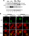
Interaction of Nup98/HoxA9 with Rae1 is similar to that of Nup98. HeLa cells were transfected and synchronized in M phase when indicated. U, unsynchronized cells. (A) Lysates were prepared from equivalent numbers of interphase or mitotic cells, immunoprecipitated using anti-GFP, and equivalent amounts of precipitate were immunoblotted using anti-GFP or anti-Rae1. The presence of shifted bands (dots, lane 6) due to phosphorylation of Nup98 confirms the mitotic nature of lysates from the synchronized cells. Similar amounts of Rae1 are immunoprecipitated with both GFP-Nup98 and GFP-Nup98/HoxA9. (B) Transfected cells expressing GFP-Nup98 (a, c, e, g, i, and k) or GFP-Nup98/HoxA9 (b, d, f, h, j, and l) were synchronized in M phase when indicated and fixed for immunofluorescence with anti-Rae1 (red). The Rae1 localization pattern is equivalent in the presence of either form of Nup98. Scale bars, 5 μm.
Figure 7.
Figure 7.
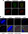
Nup98/HoxA9 is associated with the outer kinetochore during mitosis. HeLa cells were transfected, synchronized in M phase and fixed for immunofluorescence. (A) CREST serum was used to mark the inner kinetochores. Wide-field images were deconvolved using Simple PCI software. (a–c) GFP-Nup98 fusions (green) expressed at moderate levels were found in a punctate pattern along the chromosomes. CREST staining is shown in red. (d–f) When expressed at very low levels, GFP-Nup98/HoxA9 is found only at kinetochores during mitosis, overlapping or adjacent to CREST antigens. Scale bar, 5 μm. (B) HeLa cells expressing a low level of GFP-Nup98 (green) were fixed and stained for DNA (Hoechst, blue, a and f and inset in h), CenpA (red), and CREST (blue, d, e, g, and h). Arrowheads indicate unattached kinetochores positive for Nup98/HoxA9. (a–e) Deconvolved images from a single plane of a z-series. (f–h) Surface renderings of the entire z-stack. GFP-Nup98/HoxA9 (green) is found immediately adjacent to CenpA (red). The inset in panel h shows the DNA stain added to the image in the panel.

Similar articles

Cited by

References

    1. Argiropoulos B., Humphries R. K. Hox genes in hematopoiesis and leukemogenesis. Oncogene. 2007;26:6766–6776. - PubMed
    1. Babu J. R., Jeganathan K. B., Baker D. J., Wu X., Kang-Decker N., Van Deursen J. M. Rae1 is an essential mitotic checkpoint regulator that cooperates with Bub3 to prevent chromosome missegregation. J. Cell Biol. 2003;160:341–353. - PMC - PubMed
    1. Bai X. T., Gu B. W., Yin T., Niu C., Xi X. D., Zhang J., Chen Z., Chen S. J. Trans-repressive effect of NUP98-PMX1 on PMX1-regulated c-FOS gene through recruitment of histone deacetylase 1 by FG repeats. Cancer Res. 2006;66:4584–4590. - PubMed
    1. Blevins M. B., Smith A. M., Phillips E. M., Powers M. A. Complex formation among the RNA export proteins Nup98, Rae1/Gle2, and TAP. J. Biol. Chem. 2003;278:20979–20988. - PubMed
    1. Brown J. A., Bharathi A., Ghosh A., Whalen W., Fitzgerald E., Dhar R. A mutation in the Schizosaccharomyces pombe rae1 gene causes defects in Poly(A+) RNA export and in the cytoskeleton. J. Biol. Chem. 1995;270:7411–7419. - PubMed

Publication types

MeSH terms