. 2010 May 1;21(9):1629-42.
doi: 10.1091/mbc.e09-09-0809. Epub 2010 Mar 17.

Endogenous RhoG is rapidly activated after epidermal growth factor stimulation through multiple guanine-nucleotide exchange factors

Affiliations

Endogenous RhoG is rapidly activated after epidermal growth factor stimulation through multiple guanine-nucleotide exchange factors

Thomas Samson et al. Mol Biol Cell. .

Erratum in

  • Mol Biol Cell. 2011 Jul 15;22(14):2659

Abstract

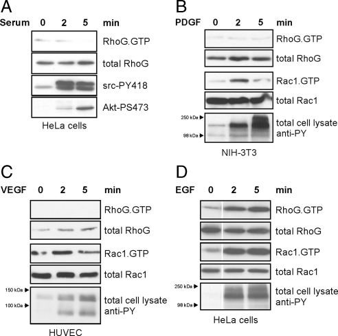
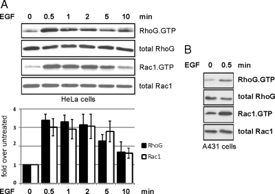
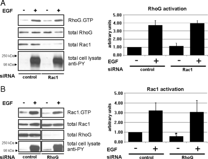
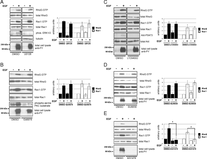
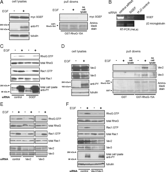
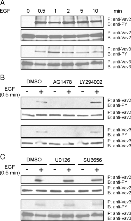
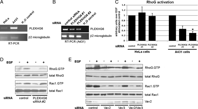
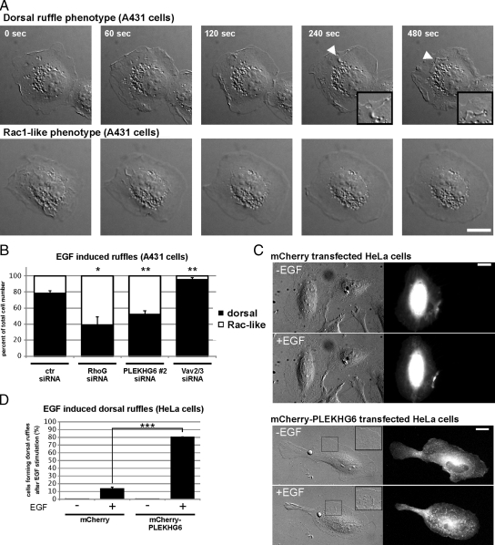
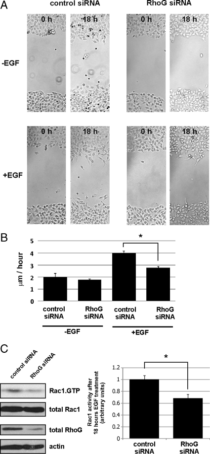
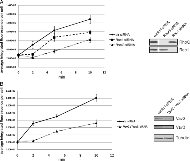
RhoG is a member of the Rac-like subgroup of Rho GTPases and has been linked to a variety of different cellular functions. Nevertheless, many aspects of RhoG upstream and downstream signaling remain unclear; in particular, few extracellular stimuli that modulate RhoG activity have been identified. Here, we describe that stimulation of epithelial cells with epidermal growth factor leads to strong and rapid activation of RhoG. Importantly, this rapid activation was not observed with other growth factors tested. The kinetics of RhoG activation after epidermal growth factor (EGF) stimulation parallel the previously described Rac1 activation. However, we show that both GTPases are activated independently of one another. Kinase inhibition studies indicate that the rapid activation of RhoG and Rac1 after EGF treatment requires the activity of the EGF receptor kinase, but neither phosphatidylinositol 3-kinase nor Src kinases. By using nucleotide-free RhoG pull-down assays and small interfering RNA-mediated knockdown studies, we further show that guanine-nucleotide exchange factors (GEFs) of the Vav family mediate EGF-induced rapid activation of RhoG. In addition, we found that in certain cell types the recently described RhoG GEF PLEKHG6 can also contribute to the rapid activation of RhoG after EGF stimulation. Finally, we present results that show that RhoG has functions in EGF-stimulated cell migration and in regulating EGF receptor internalization.


Figures

Figure 1.
Figure 1.
RhoG is activated after EGF stimulation but not by other growth factors or serum. Serum-starved cells were treated with the indicated growth factors or serum for 0, 2, and 5 min. Endogenous RhoG and Rac1 activities were measured from total cell lysates by pull-down assays by using GST-ELMO and GST-PBD, respectively. (A) HeLa cells were treated with 20% serum. Confirmation that Src and PI3K signaling occurred in responses to the serum treatment was done by blotting total cell lysates for Src-PY418 and Akt-PS473. (B) NIH-3T3 fibroblasts were treated with 20 ng/ml PDGF. (C) HUVECs were treated with 20 ng/ml VEGF. (D) HeLa cells were treated with EGF (20 ng/ml). Cellular responses in B, C, and D to stimulation by the specific growth factors were measured by blotting total cell lysates for phospho-tyrosine at the molecular weights corresponding to the relevant receptor tyrosine kinases.
Figure 2.
Figure 2.
EGF induces rapid activation of RhoG and Rac1 in different cell types within seconds. Endogenous levels of RhoG.GTP and Rac1.GTP were measured in serum-starved HeLa cells (A) or A431 cells (B) after EGF treatment (20 ng/ml) for the indicated times. The bar graph summarizes multiple independent experiments with HeLa cells (n ≥ 3; error bars represent SEM).
Figure 3.
Figure 3.
Rapid activation of RhoG and Rac1 after EGF treatment is not interdependent. (A and B) HeLa cells were transfected with the indicated siRNAs: Rac1-specific siRNA (A), RhoG-specific siRNA (B), and control siRNA (A and B). Rapid activation of RhoG or Rac1 after EGF treatment was measured using GST-ELMO and GST-PBD pull-down assays, respectively. The bar graphs summarize the results of multiple independent experiments (n ≥ 3; error bars represent SEM). The asterisk indicates a significant difference (p < 0.05) in basal Rac1 activity between nonstimulated control siRNA and nonstimulated RhoG siRNA-transfected cells.
Figure 4.
Figure 4.
Effects of different kinase inhibitors on rapid activation of RhoG and Rac1 after EGF treatment. Serum-starved HeLa cells were stimulated with EGF for 30 s in the presence of different pharmacological inhibitors as indicated (pretreatment for 1 h). (A) MEK1/2 inhibitor, U0126 (10 nM). (B) PKCα inhibitor, Gö6976 (1.32 μM). (C) PI3K inhibitor, LY294002 (30 nM). (D) Src-family kinase inhibitor, SU6656 (2.5 nM). (E) EGFR kinase inhibitor, AG1478 (10 μM). RhoG.GTP and Rac1.GTP were measured by pull-down assays. The bar graphs summarize multiple independent experiments (n ≥ 3; error bars represent SEM). The asterisks indicate significant differences (p < 0.05).
Figure 5.
Figure 5.
GEFs of the Vav family mediate rapid activation of RhoG and Rac1. (A) HeLa cells overexpressing myc-SGEF were lysed without stimulation or after 30 s of EGF treatment (20 ng/ml). The fraction of active SGEF was precipitated using GST-RhoG-15A protein and revealed by Western blotting. (B) Knockdown efficiency of SGEF siRNA was tested by SGEF-specific RT-PCR from control- and SGEF-siRNA–transfected HeLa cells. RT-PCR of β2-microglobulin RNA served as a control. (C) HeLa cells were transfected 72 h before EGF stimulation with control or SGEF-specific siRNA. Rapid activation of RhoG or Rac1 after 30 s of EGF treatment was measured using ELMO- and PBD-pull-down assays, respectively. (D) Serum-starved HeLa cells were lysed without any stimulus or after 30 s of EGF treatment (20 ng/ml) and activated RhoG-specific GEFs were precipitated using GST-RhoG-15A protein. Immunoblots of the precipitates revealed that both Vav2 and Vav3 are rapidly activated in HeLa cells after EGF stimulation (right). (E and F) Experimental conditions as in B; siRNAs: control, Vav2, and Vav3 (E); control, Vav2/Vav3 simultaneously (F).
Figure 6.
Figure 6.
Rapid phosphorylation of Vav2 and Vav3 after EGF stimulation. HeLa cells were stimulated with EGF (20 ng/ml) for the indicated times. Immunoprecipitated Vav2 and Vav3 were blotted for phospho-tryosine and Vav2 or Vav3, respectively. (A) Kinetics of Vav2 and Vav3 phosphorylation after different times of EGF stimulation. (B and C) Preceding EGF stimulation for 30 s, the cells were treated for 1 h with pharmacological inhibitors for different kinases: EGFR kinase inhibitor: AG1478 (10 nM); PI3K inhibitor, LY294002 (30 nM); MEK1/2 inhibitor, U0126 (10 nM); Src-family kinase inhibitor, SU6656 (2.5 nM).
Figure 7.
Figure 7.
PLEKHG6 and Vav-family GEFs are involved in EGF stimulated RhoG activation in A431 cells. (A) RT-PCR analysis with primers specific for PLEKHG6 was performed with total RNA samples from HeLa and A431 cells. RT-PCR of β2-microglobulin RNA served as a control. (B) To confirm knockdown with the PLEKHG6 siRNAs, RT-PCR analysis with primers specific for PLEKHG6 was performed with total RNA samples from A431 cells that have been transfected with control or one of two different PLEKHG6-specific siRNAs (PLEKHG6 #1, PLEKHG6 #2). RT-PCR of β2-microglobulin RNA served as a control. (C) A431 cells and HeLa cells were transfected with control siRNA or one of two different PLEKHG6-specific siRNAs (PLEKHG6 #1, PLEKHG6 #2). All cells were stimulated for 30 s with EGF before GST-ELMO pull-down assays were performed. The bar graphs represent RhoG activity 30 s after EGF treatment in PLEKHG6 knockdown conditions compared with siRNA controls (n = 3; error bars represent SEM). The asterisk indicates significant differences compared with control siRNA-transfected cells (p < 0.05). (D and E) A431 cells were transfected with the indicated siRNAs: control, PLEKHG6 #2 (D); control, Vav2, Vav3, and Vav2/Vav3 (E). Rapid activation of RhoG or Rac1 after 30 s EGF treatment was measured using GST-ELMO and GST-PBD pull-down assays, respectively.
Figure 8.
Figure 8.
RhoG and PLEKHG6 are required for dorsal ruffle formation in A431 cells after EGF stimulation. A431 cells were transfected with control, RhoG-, or PLEKHG6-specific siRNAs. After 72 h, the cells were plated on fibronectin-coated coverslips (5 μg/ml). After 4 h of serum starvation, the cells were treated with 20 ng/ml EGF and DIC microscopy movies of individual cells were taken (10 s/frame; 480 s total). Cellular ruffling responses showed either a “dorsal ruffling phenotype” (A, top row, arrowheads indicate dorsal ruffles, also shown enlarged in the inset) or a “Rac-like ruffling phenotype” (A, bottom row). Bar, 20 μm. (B) Quantification of the occurrence of dorsal-ruffle phenotype versus Rac-like phenotype after transfecting the different siRNAs. Shown are averaged results from three independently performed experiments. Asterisks indicate significant difference compared with control siRNA transfected cells (Student's t test, p < 0.05 and p < 0.01, respectively). (C) HeLa cells were transfected with low amounts of expression constructs for mCherry or mCherry-PLEKHG6 and analyzed for dorsal ruffle formation after EGF treatment. Bars, 20 μm. (D) Quantification of the frequency of EGF induced dorsal ruffle formation of HeLa cells transfected with the indicated expression constructs. Shown are averaged results from three independently performed experiments. Asterisks indicate significant differences (Student's t test, p < 0.001).
Figure 9.
Figure 9.
EGF-induced cell migration is regulated by RhoG. HeLa cells were transfected with the indicated siRNAs. The cell monolayers were starved before wounding and cultured with or without EGF. (A) Representative images of wounds immediately after wounding (0 h) or after 18 h. (B) Averaged migration speed of the cell fronts calculated from 36 image sets per condition (0 h/18 h) as described in Materials and Methods. Asterisks indicate significant differences between control and RhoG siRNA-transfected cells (Student's t test, p < 0.05). (C) Rac1 activity was determined in control or RhoG siRNA-transfected HeLa cells that were exposed to EGF containing media for 18 h. The bar graph on the right summarizes three different experiments. Asterisks indicate significant differences between control and RhoG siRNA-transfected cells (Student's t test, p < 0.05).
Figure 10.
Figure 10.
EGFR internalization is modulated by RhoG and Rac1. HeLa cells were transfected with the indicated siRNAs 72 h before the experiment (A: RhoG or Rac1; B: Vav2/Vav3). After binding of EGF-Alexa488 (1 μg/ml in PBS) for 1 h at 4°C to HeLa cells, EGFR internalization was induced by shifting the temperature to 37°C for the indicated times. The integrated fluorescence intensities of multiple cells from each transfection were measured and averaged. Error bars represent SEM. All measured time points were significantly different from the control (p < 0.001), except at 0 min.

Similar articles

Cited by

References

    1. Araki N., Egami Y., Watanabe Y., Hatae T. Phosphoinositide metabolism during membrane ruffling and macropinosome formation in EGF-stimulated A431 cells. Exp. Cell Res. 2007;313:1496–1507. - PubMed
    1. Beier I., Dusing R., Vetter H., Schmitz U. Epidermal growth factor stimulates Rac1 and p21-activated kinase in vascular smooth muscle cells. Atherosclerosis. 2008;196:92–97. - PubMed
    1. Bertrand-Duchesne M. P., Grenier D., Gagnon G. Epidermal growth factor released from platelet-rich plasma promotes endothelial cell proliferation in vitro. J. Periodontal Res. 2010;45:87–93. - PubMed
    1. Bishop A. L., Hall A. Rho GTPases and their effector proteins. Biochem. J. 2000;348:241–255. - PMC - PubMed
    1. Blangy A., Vignal E., Schmidt S., Debant A., Gauthier-Rouviere C., Fort P. TrioGEF1 controls Rac- and Cdc42-dependent cell structures through the direct activation of rhoG. J. Cell Sci. 2000;113:729–739. - PubMed

Publication types

MeSH terms