. 2010 Aug;27(8):1923-34.
doi: 10.1093/molbev/msq080. Epub 2010 Mar 17.

Accelerated evolution of PAK3- and PIM1-like kinase gene families in the zebra finch, Taeniopygia guttata

Affiliations

Accelerated evolution of PAK3- and PIM1-like kinase gene families in the zebra finch, Taeniopygia guttata

Lesheng Kong et al. Mol Biol Evol. 2010 Aug.

Abstract

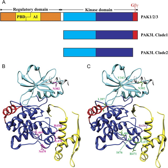
Genes encoding protein kinases tend to evolve slowly over evolutionary time, and only rarely do they appear as recent duplications in sequenced vertebrate genomes. Consequently, it was a surprise to find two families of kinase genes that have greatly and recently expanded in the zebra finch (Taeniopygia guttata) lineage. In contrast to other amniotic genomes (including chicken) that harbor only single copies of p21-activated serine/threonine kinase 3 (PAK3) and proviral integration site 1 (PIM1) genes, the zebra finch genome appeared at first to additionally contain 67 PAK3-like (PAK3L) and 51 PIM1-like (PIM1L) protein kinase genes. An exhaustive analysis of these gene models, however, revealed most to be incomplete, owing to the absence of terminal exons. After reprediction, 31 PAK3L genes and 10 PIM1L genes remain, and all but three are predicted, from the retention of functional sites and open reading frames, to be enzymatically active. PAK3L, but not PIM1L, gene sequences show evidence of recurrent episodes of positive selection, concentrated within structures spatially adjacent to N- and C-terminal protein regions that have been discarded from zebra finch PAK3L genes. At least seven zebra finch PAK3L genes were observed to be expressed in testis, whereas two sequences were found transcribed in the brain, one broadly including the song nuclei and the other in the ventricular zone and in cells resembling Bergmann's glia in the cerebellar Purkinje cell layer. Two PIM1L sequences were also observed to be expressed with broad distributions in the zebra finch brain, one in both the ventricular zone and the cerebellum and apparently associated with glial cells and the other showing neuronal cell expression and marked enrichment in midbrain/thalamic nuclei. These expression patterns do not correlate with zebra finch-specific features such as vocal learning. Nevertheless, our results show how ancient and conserved intracellular signaling molecules can be co-opted, following duplication, thereby resulting in lineage-specific functions, presumably affecting the zebra finch testis and brain.


Figures

F<sc>IG</sc>. 1.
FIG. 1.
Phylogenetic tree of PAK-related genes constructed using MrBayes 3.1.2 (Ronquist and Huelsenbeck 2003). Sequences are derived from budding yeast (Saccharomyces cerevisiae): Ste20 (Ensembl ID: YHL007C); human (Homo sapiens): PAK1 (ENSG00000149269), PAK2 (ENSG00000180370), PAK3 (ENSG00000077264), PAK4 (ENSG00000130669), PAK6 (ENSG00000137843), and PAK7 (ENSG00000101349); chicken (Gallus gallus): PAK1 (ENSGALG00000000681), PAK2 (ENSGALG00000006426), PAK3 (ENSGALG00000008058), PAK4 (ENSGALG00000014303), PAK6 (ENSGALG00000004827), and PAK7 (ENSGALG00000008947); zebra finch (Taeniopygia guttata): PAK1 (ENSTGUG00000013034), PAK2 (ENSTGUG00000005832), PAK3 (ENSTGUG00000004384), PAK6 (ENSTGUG00000007255), PAK7 (ENSTGUG00000006182), PAK3L-1 (ENSTGUG00000000383), PAK3L-2 (ENSTGUG00000001058), PAK3L-3 (ENSTGUG00000001397), PAK3L-4 (ENSTGUG00000006253), PAK3L-5 (ENSTGUG00000006415), PAK3L-6 (ENSTGUG00000013873), PAK3L-7 (ENSTGUG00000014676), PAK3L-8 (ENSTGUG00000014731), PAK3L-9 (ENSTGUG00000015007), PAK3L-10 (ENSTGUG00000015265), PAK3L-11 (ENSTGUG00000016297), PAK3L-12 (ENSTGUG00000016405), PAK3L-13 (ENSTGUG00000016760), PAK3L-14 (ENSTGUG00000016946), PAK3L-15 (ENSTGUG00000017512), PAK3L-16 (ENSTGUG00000017514), PAK3L-17 (ENSTGUG00000017522), PAK3L-18 (ENSTGUG00000017537), PAK3L-19 (ENSTGUG00000003740), PAK3L-20 (ENSTGUG00000017535), PAK3L-21 (ENSTGUG00000014645), PAK3L-22 (ENSTGUG00000014689), PAK3L-23 (ENSTGUG00000014769), PAK3L-24 (ENSTGUG00000015498), PAK3L-25 (ENSTGUG00000016398), PAK3L-26 (ENSTGUG00000017061), PAK3L-27 (ENSTGUG00000017214), PAK3L-28 (ENSTGUG00000013955), PAK3L-29 (ENSTGUG00000014357), PAK3L-30, and PAK3L-31. Nucleotide sequences were used to estimate the tree under the following parameters: GTR+I+G, 5 million generations, 4 × 4 nucleotide substitution, sampled every 100 generations, with the consensus tree drawn using the last 25 000 trees. PAK4, PAK6, and PAK7 genes and the yeast Ste20 gene were used as outgroup sequences and are not shown in this figure. The shaded rectangle with solid lines delineates the clade of all zebra finch PAK3L genes. Similarly, the dashed rectangle delineates Clade 1 and the dotted rectangle Clade 2. The estimated formula image 
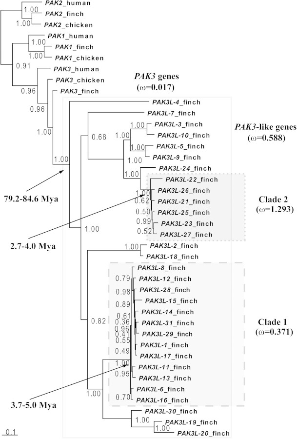
(nonsynonymous/synonymous substitution rates ratio) and divergence times (in Mya) for the clade including all PAK3L genes, Clades 1 and 2 are shown.
F<sc>IG</sc>. 2.
FIG. 2.
Brain expression of PAK3 and PAK3L genes in adult male zebra finches revealed by in situ hybridization. (A and B) PAK3 has a broad distribution, without major regional specializations; expression in all major song control nuclei is similar to that in the surrounding regions. Shown are parasagittal sections at ∼0.8 (A) and 2.0 (B) mm from the midline. (CI) Expression of clone DV950892 is restricted to the ventricular zone and cerebellum. (C) Diagram of parasagittal section at ∼1.4 mm indicating position of the photomicrographs in (DI). (D) High expression in the caudal-most portion of the lateral ventricular zone. (E) Expression is high in the Purkinje cell layer (pcl) and lower in the granule cell layer (gcl) and molecular layer (ml) of the cerebellum. (F) High expression in the rostral-most portion of the lateral ventricular zone (black arrowheads) but not in the intermediate portion (white arrowheads). (G) Detailed view of the ventricular zone shows high expression in the ependymal lining. (H) Detailed view of the same region as in G, taken from an adjacent Nissl-stained section shows the high cell density in the ventricular zone. (I) Detailed view of the cerebellum shows expression in the small glial-like cells but not in the large Purkinje cells (indicated by asterisks) of the pcl; expression is much lower in the gcl and absent in the ml. Scale bars = 100 μm in (DF) and 10 μm in (GI). A, arcopallium; Cb, cerebellum; CMM, caudomedial mesopallium; DM, dorsomedial nucleus of the intercollicular complex; gcl, granule cell layer of the cerebellum; H, hyperpallium; Hp, hippocampus; HVC, vocal control nucleus HVC (nucleus hyperstriatalis ventralis, pars caudalis); Hypo, hypothalamus; LMAN, lateral part of the magnocellular nucleus of the anterior nidopallium; M, mesopallium; ml, molecular layer of the cerebellum; N, nidopallium; NCM, caudomedial nidopallium; pcl, Purkinje cell layer of the cerebellum; RA, robust nucleus of the arcopallium; St, striatum; Tel, telencephalon; TeO, optic tectum; Thal, thalamus; X, area X of the striatum.
F<sc>IG</sc>. 3.
FIG. 3.
Schematic representation of PAK domain structures and positively selected amino acid sites in zebra finch PAK3L genes mapped on to human PAK1 structure. (A) Schematic representation of PAK1/2/3, PAK3L Clade 1 and Clade 2. The autoregulatory segment is shown in yellow, which consists of the p21-binding domain (PBD) and the autoinhibitory domain (AI). For the kinase domain, the N lobe is in light blue and C lobe is in dark blue. The putative Gβγ-binding motif (corresponding to exon 8 of PAK3L genes) is in red. The α-helix in the N-terminal region is in white (this region is absent in PAK3L genes). (B) and (C) show different sets of positively selected amino acid sites in stick form and labeled: (B) site analysis for all PAK3L genes (positively selected sites are colored in pink); (C) Clade-wise site analysis (positively selected sites from Clade 1 are in purple and sites from Clade 2 in green). Figures were produced using the UCSF Chimera package (Pettersen et al. 2004).
F<sc>IG</sc>. 4.
FIG. 4.
Phylogenetic tree of PIM-related genes constructed using PHYML v3.0.1 (Guindon and Gascuel 2003). Nucleotide sequences are derived from nematode (Caenorhabditis elegans): PIM (NCBI GenPept ID: NP_497696.2); human (Homo sapiens): PIM1 (Ensembl ID: ENSG00000137193), PIM2 (ENSG00000102096), and PIM3 (ENSG00000198355); chicken (Gallus gallus): PIM1 (ENSGALG00000000742) and PIM3 (ENSGALG00000023576); zebra finch (Taeniopygia guttata): PIM1 (ENSTGUG00000000954), PIM3 (ENSTGUG00000003897), PIM1L-1 (ENSTGUG00000016485), PIM1L-2 (ENSTGUG00000015552), PIM1L-3 (ENSTGUG00000016751), PIM1L-4 (ENSTGUG00000015140), PIM1L-5 (ENSTGUG00000013726), PIM1L-6 (ENSTGUG00000014083), PIM1L-7 (ENSTGUG00000014454), PIM1L-8 (ENSTGUG00000015232), PIM1L-9 (ENSTGUG00000015242), and PIM1L-10 (ENSTGUG00000016198). The tree was bootstrapped for 1,000 replicates, and bootstrap values are shown on nodes. PIM_nematode was used as an outgroup. The shaded rectangle delineates the zebra finch clade of PIM1L genes. The estimated formula image for PIM1L genes and other PIM1 genes are provided. The estimated timescales for the duplications of PIM1L genes are shown (in Mya).
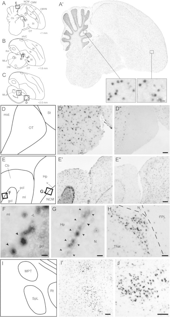
F<sc>IG</sc>. 5.
FIG. 5.
Brain expression of PIM1L genes in adult male zebra finches revealed by in situ hybridization. (AC) Diagrams of parasagittal sections depicting the brain levels analyzed for expression of PIM1L genes; squares indicate the position of the photomicrographs in (DJ). (A′) DV954383 exhibits a broad and largely uniform brain distribution, with some enrichment in the ventricular zone and cerebellum; insets show detailed views of labeling at the cellular level for the cerebellar molecular layer and the hyperpallium, with a punctate distribution that appears to be largely nuclear. (D) Schematic diagram depicting portions of the midbrain and optic tract (location indicated in C) shown in (D′) and (D′′). (D′) DV954383 is uniformly expressed in a discrete cell population in the optic tract and in adjacent parts of the midbrain. (D′′) DV959014 is expressed in the midbrain but is clearly absent in the optic tract. (E) Schematic diagram depicting portions of the cerebellum and hippocampus (location indicated in A) shown in (E′) and (E′′). (E′) DV954383 is strongly expressed in the Purkinje cell layer and in sparse cell populations in the molecular and granular cell layers of the cerebellum. (E′′) DV959014 has uniform and moderate expression in the hippocampus and NCM but lacks enrichment in different layers of the cerebellum or in the lateral ventricle. (F) Detailed view of the cerebellum (location shown in E) demonstrates strong expression of DV954383 over some Purkinje cells (black arrowheads), whereas others are clearly negative (empty arrowhead), and over discrete cells in the molecular layer. (G) Detailed view of the lateral ventricle (location shown in E) demonstrates strong expression of DV954383 over discrete cells in the ventricular zone (indicated by black arrowheads) between the hippocampus and the nidopallium. (H) Detailed view of the dorsal thalamus (location shown in B) demonstrates strong expression of DV959014 in a discrete nucleus of unknown identity. (I) Schematic diagram depicting portions of the thalamus and pretectal area (location indicated in C) shown in (I′). (I′) DV954383 shows enriched expression in the medial pretectal nucleus and in nucleus spiriformis lateralis. (J) DV959014 is highly enriched in the nucleus spiriformis medialis. Scale bars: 100 μm for (D), (E), (H), (I), and (J); 10 μm for (F) and (G), and for insets in (A′). Abbreviations: A, arcopallium; Cb, cerebellum; CMM, caudomedial mesopallium; DM, dorsomedial nucleus of the intercollicular complex; FPL, fasciculus prosencephali lateralis; gcl, granule cell layer of the cerebellum; H, hyperpallium; Hp, hippocampus; HVC, vocal control nucleus HVC (nucleus hyperstriatalis ventralis, pars caudalis); LMAN, lateral part of the magnocellular nucleus of the anterior nidopallium; M, mesopallium; mid, midbrain; ml, molecular layer of the cerebellum; MLd, dorsal part of the lateral mesencephalic nucleus; MPT, medial pretectal nucleus; N, nidopallium; NCM, caudomedial nidopallium; OT, optic tract; pcl, Purkinje cell layer of the cerebellum; RA, robust nucleus of the arcopallium; Rt, nucleus rotundus; Sp, nucleus spiriformis; St, striatum; Tel, telencephalon; TeO, optic tectum; Thal, thalamus; v, ventricle; X, area X of the striatum.

Similar articles

Cited by

References

    1. Allen KM, Gleeson JG, Bagrodia S, Partington MW, MacMillan JC, Cerione RA, Mulley JC, Walsh CA. PAK3 mutation in nonsyndromic X-linked mental retardation. Nat Genet. 1998;20:25–30. - PubMed
    1. Altschul SF, Gish W, Miller W, Myers EW, Lipman DJ. Basic local alignment search tool. J Mol Biol. 1990;215:403–410. - PubMed
    1. Bokoch GM. Biology of the p21-activated kinases. Annu Rev Biochem. 2003;72:743–781. - PubMed
    1. Brown JW, Rest JS, Garcia-Moreno J, Sorenson MD, Mindell DP. Strong mitochondrial DNA support for a Cretaceous origin of modern avian lineages. BMC Biol. 2008;6:6. - PMC - PubMed
    1. Church DM, Goodstadt L, Hillier LW, et al. Lineage-specific biology revealed by a finished genome assembly of the mouse. PLoS Biol. 2009;7:e1000112. - PMC - PubMed

Publication types

MeSH terms

Substances