Review
. 2010 Aug;31(4):544-77.
doi: 10.1210/er.2009-0023. Epub 2010 Mar 17.

The neurobiology of preovulatory and estradiol-induced gonadotropin-releasing hormone surges

Affiliations
Review

The neurobiology of preovulatory and estradiol-induced gonadotropin-releasing hormone surges

Catherine A Christian et al. Endocr Rev. 2010 Aug.

Abstract

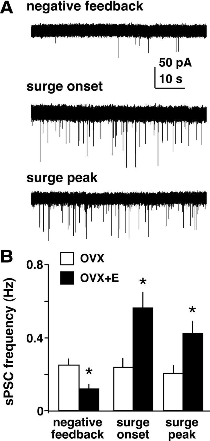
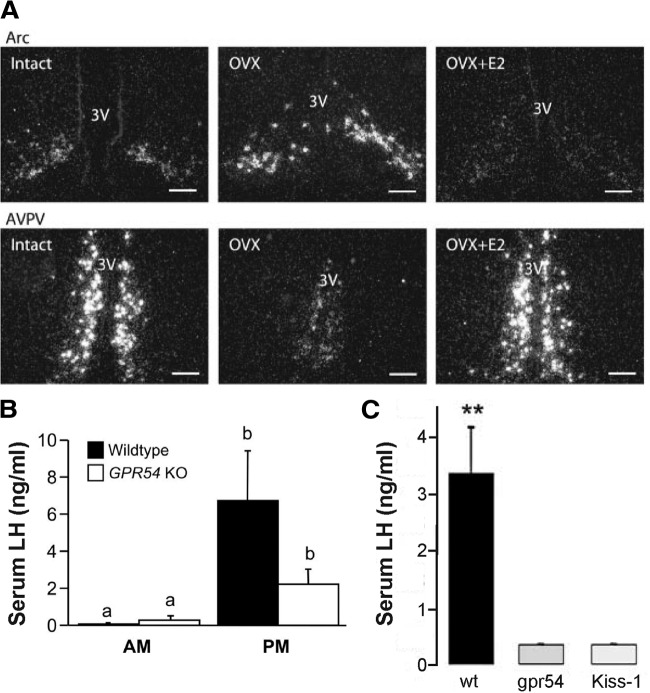
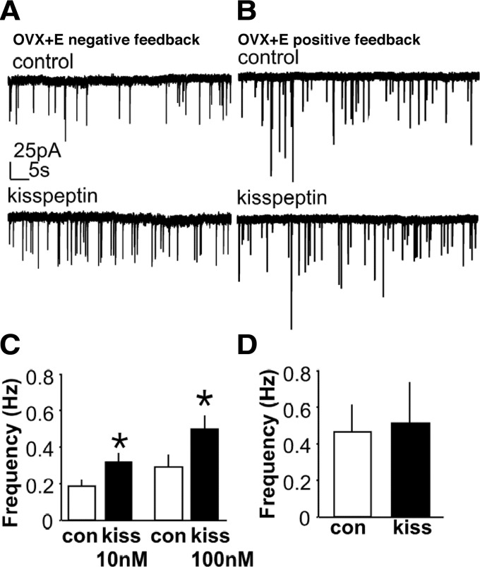
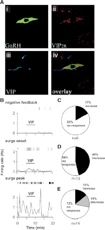
Ovarian steroids normally exert homeostatic negative feedback on GnRH release. During sustained exposure to elevated estradiol in the late follicular phase of the reproductive cycle, however, the feedback action of estradiol switches to positive, inducing a surge of GnRH release from the brain, which signals the pituitary LH surge that triggers ovulation. In rodents, this switch appears dependent on a circadian signal that times the surge to a specific time of day (e.g., late afternoon in nocturnal species). Although the precise nature of this daily signal and the mechanism of the switch from negative to positive feedback have remained elusive, work in the past decade has provided much insight into the role of circadian/diurnal and estradiol-dependent signals in GnRH/LH surge regulation and timing. Here we review the current knowledge of the neurobiology of the GnRH surge, in particular the actions of estradiol on GnRH neurons and their synaptic afferents, the regulation of GnRH neurons by fast synaptic transmission mediated by the neurotransmitters gamma-aminobutyric acid and glutamate, and the host of excitatory and inhibitory neuromodulators including kisspeptin, vasoactive intestinal polypeptide, catecholamines, neurokinin B, and RFamide-related peptides, that appear essential for GnRH surge regulation, and ultimately ovulation and fertility.


Figures

Figure 1
Figure 1
Measurement of GnRH, LH, and circulating estradiol (E2) in an individual ewe during the follicular phase demonstrates a preovulatory GnRH/LH surge. Note that the termination of the LH surge precedes that of the GnRH surge. [Data adapted from S. M. Moenter et al.: Endocrinology 129:1175–1182 (9). © 1991, The Endocrine Society].
Figure 2
Figure 2
Constant estradiol treatment of OVX mice induces daily LH surges. A, Serum LH levels (mean ± sem) sampled in OVX+E mice at 0700 h (open bars) or 1600 h (filled bars) (lights off at 1630 h). B, OVX mice show no diurnal difference in serum LH levels; estradiol administered from the time of OVX induces negative feedback in the morning and positive feedback in the evening on d 2 after surgery. *P < 0.05; ***P < 0.001; #P < 0.01 vs. d 2 p.m. [Data adapted from C. A. Christian et al.: Proc Natl Acad Sci USA 102:15682–15687 (49). © 2005, National Academy of Sciences, U.S.A.].
Figure 3
Figure 3
Triple label of recorded GnRH neuron from a GnRH-GFP transgenic mouse. A, GFP signal used to target recording pipette to a GnRH neuron. B, Biocytin labeling of recorded cell detected with streptavidin-Cy3. C, Immunostaining for GnRH with Cy5-conjugated secondary antibody. [Data adapted from K. J. Suter et al.: Endocrinology 141:412–419 (73). © 2000, The Endocrine Society].
Figure 4
Figure 4
GnRH neuron firing activity correlates with LH levels in the daily LH surge mouse model. Representative examples of firing patterns in GnRH neurons recorded extracellularly from OVX (left) or OVX+E (right) mice in the morning (top) or evening (bottom). Vertical lines at the top of each graph indicate timing of individual action currents recorded from an individual GnRH neuron. The frequency of action current firing is plotted in 1-min bins for each cell. Cells from OVX mice show no diurnal difference in firing properties, whereas cells from OVX+E mice show low levels of activity in the morning (negative feedback) and higher levels of activity in the evening (positive feedback). [Data adapted from C. A. Christian et al.: Proc Natl Acad Sci USA 102:15682–15687 (49). © 2005, National Academy of Sciences, U.S.A.].
Figure 5
Figure 5
Clock/Clock mutant mice fail to exhibit an LH surge on proestrus. A, Individual LH traces from wild-type (open circles) and Clock/Clock females (filled circles). White and black bars on x-axis represent times of lights on and lights off, respectively. B, Peak LH values obtained in wild-type and Clock/Clock female mice. *P < 0.01. [Data adapted from B. H. Miller et al.: Curr Biol 14:1367–1373 (20). © 2004, Elsevier].
Figure 6
Figure 6
Asymmetric activation of GnRH neurons in hamsters exhibiting split circadian behavioral rhythms of activity. A, Coronal brain section through the SCN of a behaviorally split hamster stained for cFos immunoreactivity. 3V, Third ventricle; OC, optic chiasm. Scale bar, 500 μm. B, GnRH neurons (blue stain) on side of brain in which SCN does not show cFos activation do not express cFos (white arrows). Scale bar, 50 μm. C, GnRH neurons on side of brain in which SCN shows cFos expression coexpress cFos (brown stain, black arrows). [Data adapted from H. O. de la Iglesia et al.: J Neurosci 23:7412–7414 (133). © 2003, Society for Neuroscience].
Figure 7
Figure 7
Anatomical studies indicate that inputs important for surge generation are located in AVPV. A–C, GnRH neuron-specific viral tracing shows that afferents to GnRH neurons expressing ERα are located in AVPV. A, Neurons in AVPV express GFP immunoreactivity after Bartha virus (Ba2001) viral injection into rostral preoptic area in GnRH-Cre transgenic mice, which retrogradely labels afferent inputs. Scale bar, 15 μm. B, Dual labeling for GFP (green) and ERα (red) shows that GnRH neuron afferents in AVPV express ERα. C, Schematic coronal brain maps showing GFP-immunoreactive afferent cells that did not express ERα (open stars) and GFP+ERα-immunoreactive cells (filled stars). aca, Anterior commisure; ARN, arcuate nucleus; f, fornix; MnPO, medial preoptic nucleus; ME, median eminence, PeN, periventricular nucleus; 3V, third ventricle. D, Micrographs of cFos expression in the AVPV (brightfield images) and GnRH neurons (insets) before (Di–Diii) and during (Div) an LH surge. During diestrus and on proestrus before the LH surge, GnRH neurons do not exhibit cFos, and few AVPV nuclei are cFos-positive; but during the surge, both GnRH neurons and numerous AVPV cells express cFos. Scale bars, 100 μm (brightfield images) and 10 μm (fluorescence images). [Data in panels A–C adapted from T. M. Wintermantel et al.: Neuron 52:271–280 (150); 227 2006, Elsevier. Data in panel D adapted from W. W. Le et al.: Endocrinology 140:510–519 (157); © 1999, The Endocrine Society].
Figure 8
Figure 8
GABA can depolarize and induce firing activity in adult GnRH neurons in rats and mice but inhibits other neurons. A and B, Representative on-cell recordings, in which the internal milieu is not disturbed, of action currents in a non-GnRH (A) and a GnRH (B) neuron from GnRH-GFP mice. Rapid application of a puff of 1 mm GABA inhibits firing in non-GnRH hypothalamic neurons but elicits action current firing in GnRH neurons. C, GABA-induced depolarization and action potential generation in rat GnRH neurons recorded using gramicidin perforated patch-clamp in current-clamp mode. The membrane potentials as set by current injection are indicated to the left of each trace. GABA was puff-applied for 600 msec as indicated by horizontal bars above the traces; 3 μm GABA elicited a relatively slow depolarization and generated action potential(s), and 20 μm GABA rapidly depolarized the cell and generated a single action potential at all membrane potentials examined. [Data in panels A and B adapted from R. A. DeFazio et al.: Mol Endocrinol 16:2872–2891 (223); 227 2002, The Endocrine Society. Data in panel C adapted from C. Yin et al.: J Neuroendocrinol 20:566–575 (233). © 2008, Wiley-Blackwell].
Figure 9
Figure 9
Estradiol decreases GABA transmission to GnRH neurons but increases it during the surge. A, Representative whole-cell voltage-clamp recordings of spontaneous GABAergic postsynaptic currents (sPSCs, downward deflections) in GnRH neurons from mice treated in the OVX+E daily surge model during negative feedback, surge onset, and surge peak. Recordings were performed in the presence of ionotropic glutamate receptor antagonists to isolate GABAergic currents. Note the increased frequency and amplitude of sPSCs during surge onset and peak compared with negative feedback. B, Mean ± sem of sPSC frequency in cells from OVX (open bars) and OVX+E (filled bars) mice during negative feedback, surge onset, and surge peak. *P < 0.05 vs. OVX. [Data adapted from C. A. Christian and S. M. Moenter: J Neurosci 27:1913–1921 (247). © 2007, Society for Neuroscience].
Figure 10
Figure 10
Estradiol regulates kisspeptin (KiSS-1) mRNA in the mouse AVPV and arcuate nucleus (Arc), and GPR54- and kisspeptin-null mice exhibit deficient LH surge generation. A, Dark-field micrographs showing KiSS-1 mRNA expression in the arcuate nucleus (top) and AVPV (bottom) in ovary-intact (left), OVX (middle), and OVX+E (right) mice. 3V, Third ventricle. Scale bars, 100 μm. B, Mean ± sem serum LH levels in wild-type and GPR54-null mice in the morning and evening. C, Mean ± sem serum LH levels in OVX+E+P mice in wild-type, GPR54-null, and kisspeptin-null mice. **P < 0.01. [Data in panel A adapted from J. T. Smith et al.: Endocrinology 146:3686–3692 (269); 227 2005, The Endocrine Society. Data in panel B adapted from H. M. Dungan et al.: J Neurosci 27:12088–12095 (276); 227 2007, Society for Neuroscience. Data in panel C adapted from J. Clarkson et al.: J Neurosci 28:8691–8697 (277); © 2008, Society for Neuroscience].
Figure 11
Figure 11
Kisspeptin increases GABAergic transmission to GnRH neurons during negative feedback but has no effect during positive feedback. A and B, Representative whole-cell voltage-clamp recordings of spontaneous GABAergic postsynaptic currents (sPSCs, downward deflections) in GnRH neurons from OVX+E mice during negative feedback (A) and positive feedback (B). C and D, Mean ± sem of sPSC frequency in cells from OVX+E mice during negative feedback (C) and positive feedback (D), with white bars showing control period and black bars showing kisspeptin treatment. Kisspeptin concentration in B and D = 10 nm. *P < 0.05. [Data adapted from J. Pielecka-Fortuna and S. M. Moenter: Endocrinology 151:291–300 (278). © 2010, The Endocrine Society].
Figure 12
Figure 12
GnRH neurons express VIP2 receptors, and VIP excitation of GnRH neurons is time-of-day-dependent. A, Rat GnRH neurons express VIP2 receptors. Confocal micrograph showing a GnRH neuron (Ai) in the rostral preoptic area that expresses VIP2 receptor protein (Aii) and is in close apposition to VIP (Aiii). Aiv, Overlay of Ai–Aiii. B, Representative examples of firing patterns are shown for GnRH neurons from mice treated in the OVX+E daily surge model recorded extracellularly during negative feedback (top), surge onset (middle), and surge peak (bottom). Vertical lines at the top of each graph indicate timing of individual action currents recorded from an individual GnRH neuron. The frequency of action current firing is plotted in 30-sec bins for each cell. C–E, Rate of response for OVX+E cells recorded during negative feedback (C), surge onset (D), and surge peak (E). [Data in panel A adapted from M. J. Smith et al.: Endocrinology 141:4317–4320 (292); 227 2000, The Endocrine Society. Data in panels B–E adapted from C. A. Christian and S. M. Moenter: Endocrinology 149:3130–3136 (293); © 2008, The Endocrine Society].
Figure 13
Figure 13
Activation of GnIH/RFRP cells is reduced during the LH surge in hamsters. A, Low-power micrograph of RFRP-immunoreactive cells expressing cFos in the dorsomedial hypothalamus on diestrus. B and C, RFRP-ir cells expressing cFos on proestrus during the trough (1800 h; B) and peak (2300 h; C) of expression. D, Mean percentages of RFRP-immunoreactive cells expressing cFos on diestrus and at various times on the day of proestrus. [Data adapted from E. M. Gibson et al.: Endocrinology 149:4958–4969 (374). © 2008, The Endocrine Society.]
Figure 14
Figure 14
Schematic working model of multiple signals involved in surge generation. The gray dotted box indicates portion of sagittal brain sketch magnified in schematic figure. For simplicity only, the substances with the most evidence for involvement in surge regulation are depicted; see text for full description of each neuromodulator. Excitatory inputs (green lines and text) arise from the AVPV, SCN, and medulla, mediated by kisspeptin (kiss), GABA, VIP, and catecholamines. Inhibitory inputs (red text) arising from the arcuate nucleus (ARC) and dorsomedial hypothalamus (DMH) mediated by endogenous opioid peptides (EOPs), NKB, and GnIH/RFRP-3 are reduced (dashed red line) during the surge. The SCN provides excitatory input to the AVPV and inhibitory input to the DMH at appropriate times of day to coordinate changes in downstream inputs to GnRH neurons. Sagittal brain sketch courtesy of Dr. Paul Heideman. (The College of William and Mary, Williamsburg, Virginia).

Similar articles

Cited by

References

    1. Belchetz PE, Plant TM, Nakai Y, Keogh EJ, Knobil E 1978 Hypophysial responses to continuous and intermittent delivery of hypothalamic gonadotropin-releasing hormone. Science 202:631–633 - PubMed
    1. Clarke IJ, Thomas GB, Yao B, Cummins JT 1987 GnRH secretion throughout the ovine estrous cycle. Neuroendocrinology 46:82–88 - PubMed
    1. Wildt L, Häusler A, Marshall G, Hutchison JS, Plant TM, Belchetz PE, Knobil E 1981 Frequency and amplitude of gonadotropin-releasing hormone stimulation and gonadotropin secretion in the rhesus monkey. Endocrinology 109:376–385 - PubMed
    1. Marshall JC, Griffin ML 1993 The role of changing pulse frequency in the regulation of ovulation. Hum Reprod 8:57–61 - PubMed
    1. Haisenleder DJ, Dalkin AC, Ortolano GA, Marshall JC, Shupnik MA 1991 A pulsatile gonadotropin-releasing hormone stimulus is required to increase transcription of the gonadotropin subunit genes: evidence for differential regulation of transcription by pulse frequency in vivo. Endocrinology 128:509–517 - PubMed

Publication types

MeSH terms