Comparative Study
. 2010 Mar 17;30(11):3933-46.
doi: 10.1523/JNEUROSCI.6054-09.2010.

Roles of NADPH oxidases in cisplatin-induced reactive oxygen species generation and ototoxicity

Affiliations
Comparative Study

Roles of NADPH oxidases in cisplatin-induced reactive oxygen species generation and ototoxicity

Hyung-Jin Kim et al. J Neurosci. .

Abstract

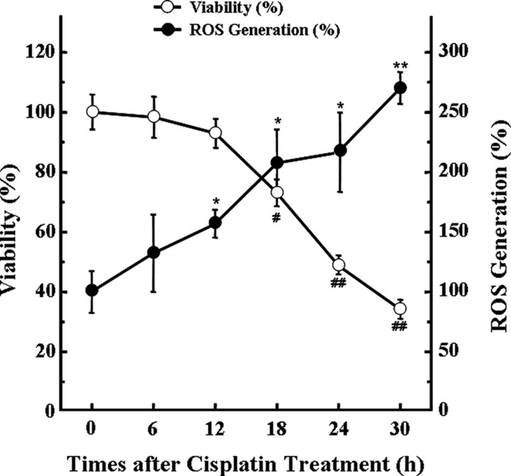
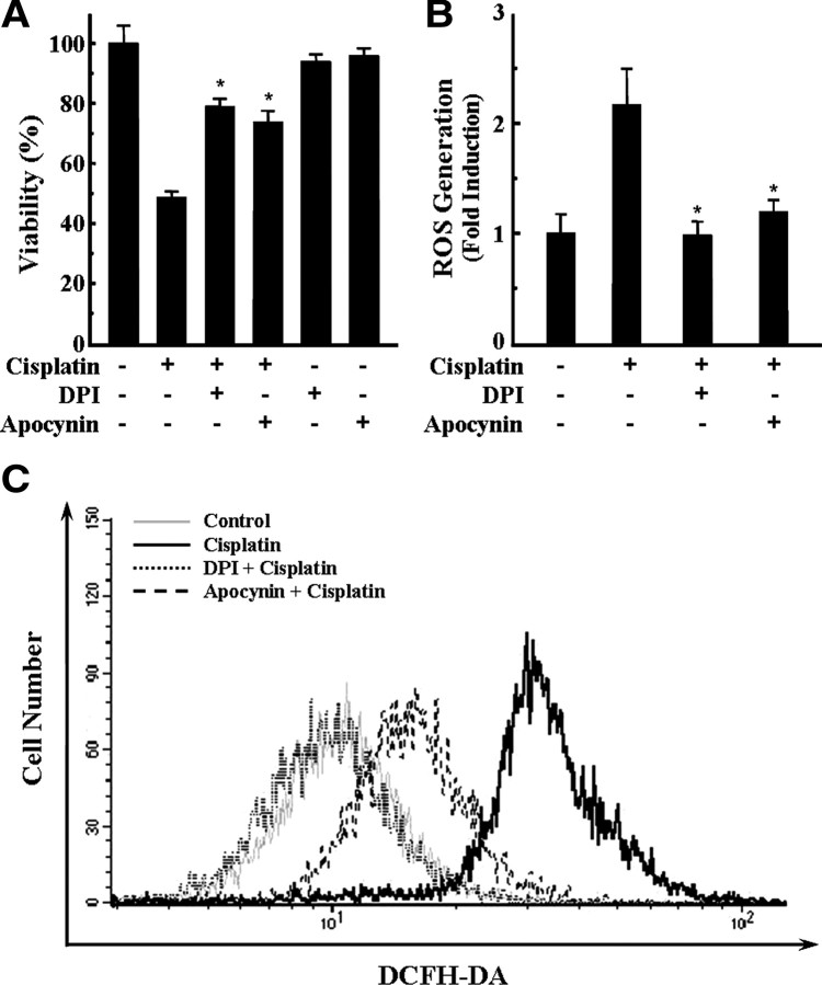
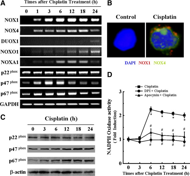
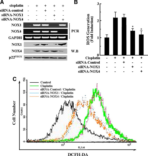
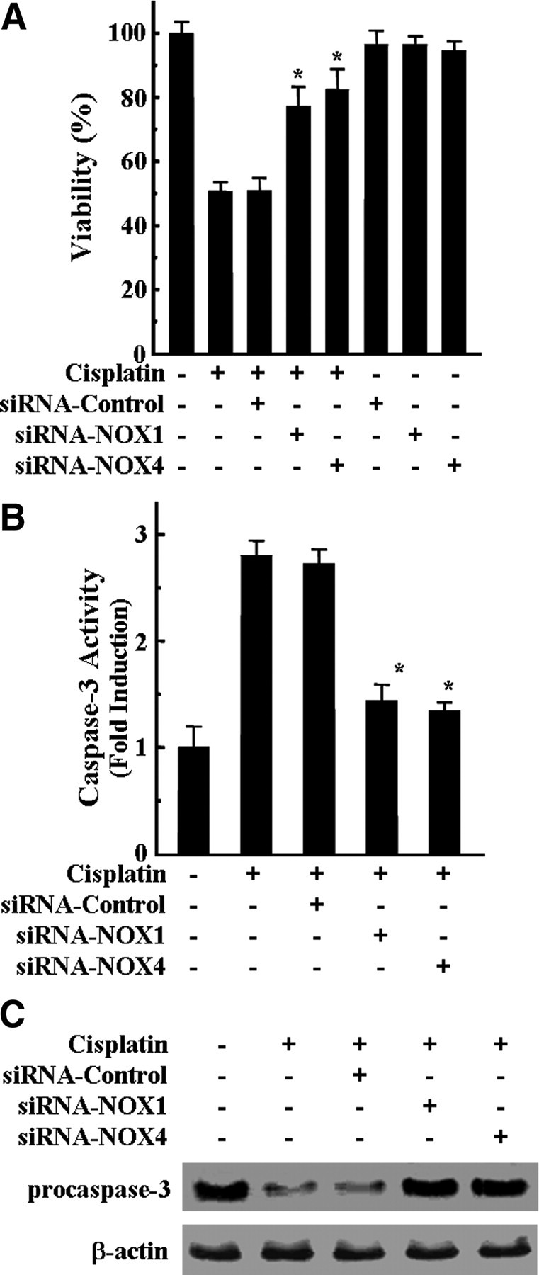
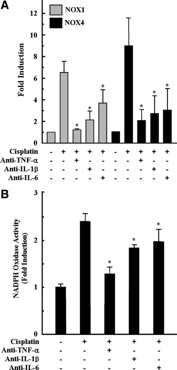
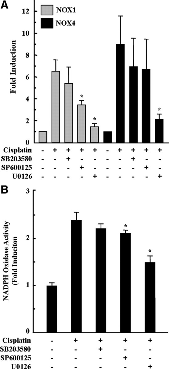
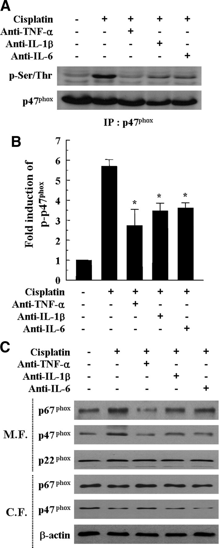
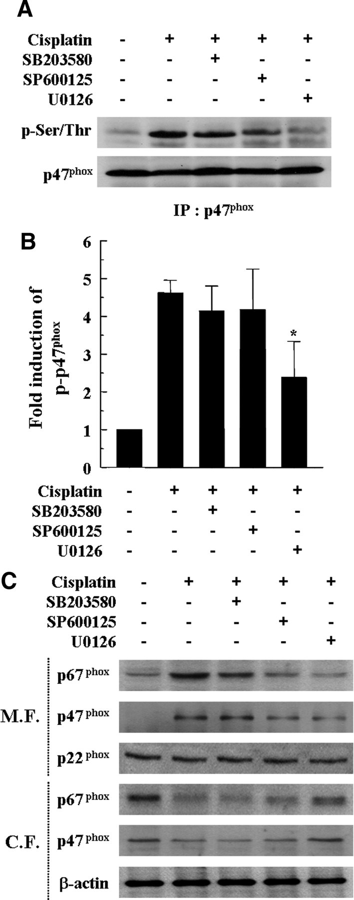
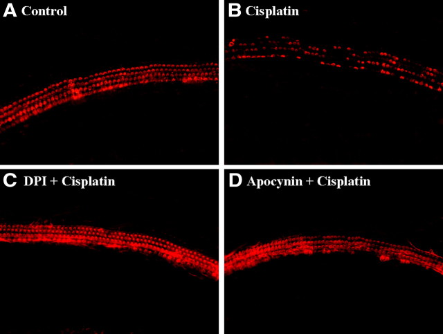
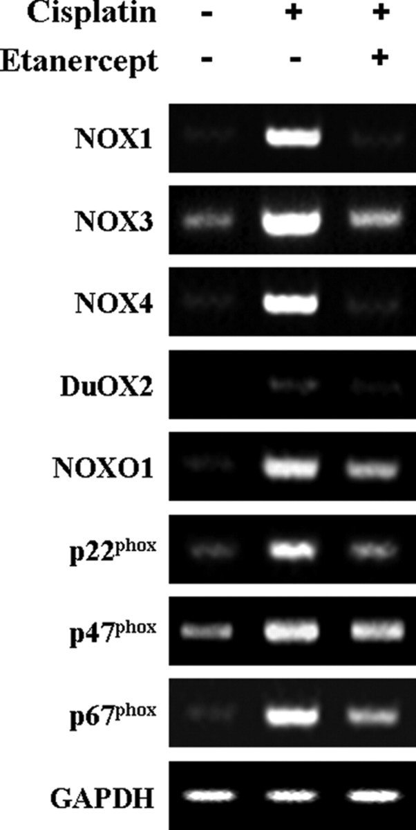
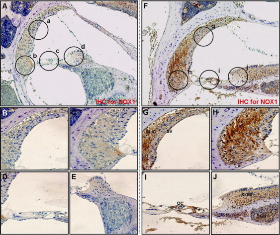
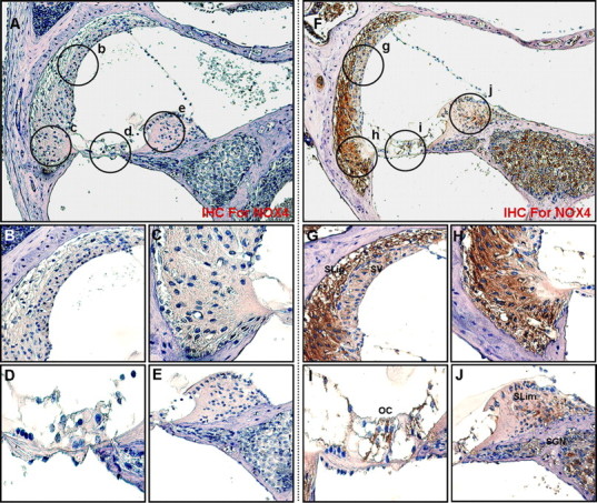
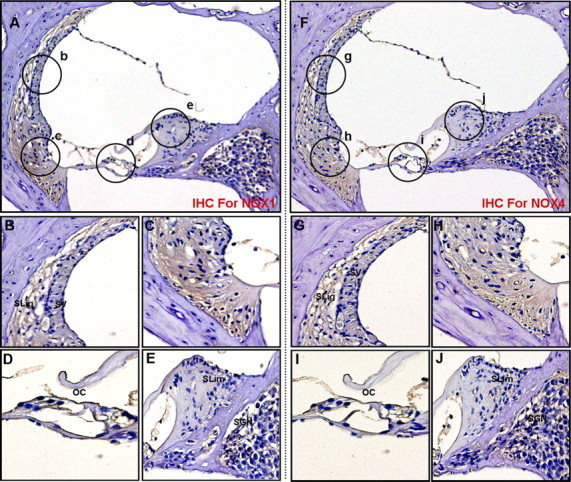
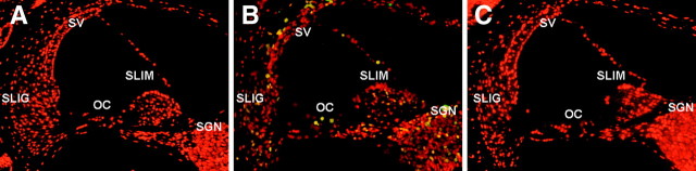
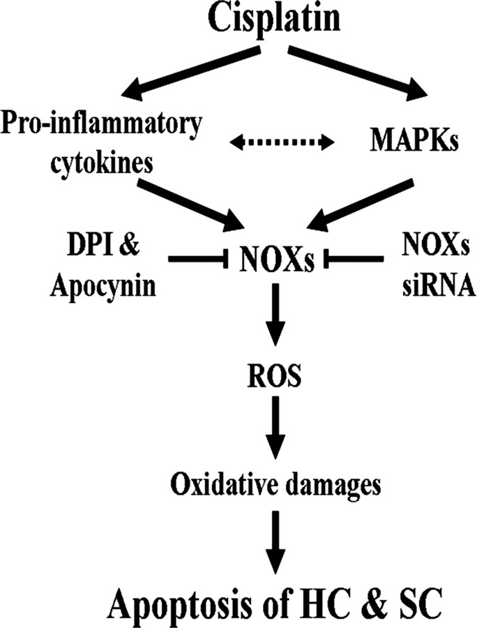
In our previous study, we clearly demonstrated the roles of pro-inflammatory cytokines, including tumor necrosis factor-alpha, interleukin-1beta (IL-1beta), and IL-6, and subsequent reactive oxygen species (ROS) generation on the pathogenesis of cisplatin ototoxicity in vitro and in vivo. ROS generation in cisplatin-treated HEI-OC1 auditory cells was also correlated with changing mitochondrial membrane potential. However, the roles of NADPH oxidase in cisplatin-induced ROS generation and ototoxicity have not been fully elucidated. Herein, immunohistochemical studies demonstrated that treatment of cisplatin induced the expression of NADPH oxidase isoforms NOX-1 and NOX-4 in HEI-OC1 auditory cells. Expression of mRNA for NOX-1, NOX-4, NOXO1, NOXA1, p47(phox), and p67(phox) was also increased. Inhibition of NADPH oxidase with diphenyleniodonium chloride or apocynin abolished ROS production and the subsequent apoptotic cell death in cisplatin-treated cells. Furthermore, suppression of NOX1 and NOX4 expression by small interfering RNA transfection markedly abolished the cytotoxicity and ROS generation by cisplatin. Together, our data suggest that ROS generated, in part, through the activation of NADPH oxidase plays an essential role in cisplatin ototoxicity.


Figures

Figure 1.
Figure 1.
Cisplatin-induced cytotoxicity is associated with an increase in ROS production in HEI-OC1 auditory cells. Cells were treated with 20 μm cisplatin for various time periods. Cell viability was then measured by MTT assay. In addition, the level of intracellular ROS was also monitored at indicated time points using a peroxide-sensitive fluorescent probe, DCFH-DA. The data represent the mean ± SD of triplicate. *,#p < 0.05, **,##p < 0.01, by one-way ANOVA compared with cell viability (*, **) or ROS generation (#, ##) in zero time (0 h) control cells.
Figure 2.
Figure 2.
Pharmacological inhibition of NADPH oxidase attenuated the cisplatin cytotoxicity and ROS generation in HEI-OC1 cells. Cells were pretreated with 100 nm DPI or apocynin, an inhibitor of NADPH oxidases, for 30 min and further incubated with 20 μm cisplatin for 24 h. A, Cell viability was then measured by MTT assay. B, After 24 h, the level of intracellular ROS was also monitored using a peroxide-sensitive fluorescent probe, DCFH-DA. The data represent the mean ± SD of three independent experiments. *p < 0.05, by one-way ANOVA compared with cisplatin-only treated cells. C, Intracellular ROS level was also measured by flow cytometry as described in Materials and Methods.
Figure 3.
Figure 3.
Various NOX isoforms are expressed in the cisplatin-treated HEI-OC1 auditory cells. A, Cells were treated with 20 μm cisplatin for the indicated time periods. Total RNA was then isolated by TRIzol, and cDNA was synthesized by reverse transcription. The cDNAs for various NOX isoforms were amplified using specific primer sets as described in Materials and Methods. B, Cells were treated with 20 μm cisplatin for 18 h. After reaction with anti-NOX1 and NOX4 antibodies as described in Materials and Methods, cells were visualized under fluorescent microscope. C, Cells were treated with 20 μm cisplatin for the indicated time periods and subjected to Western blotting with anti-p22phox, anti-p47phox, anti-p67phox, and anti-β-actin, respectively. D, Cells were treated with 20 μm cisplatin for the indicated time periods. Then, the activity of NADPH oxidase was determined using lucigenin as described in Materials and Methods. The data represent the mean ± SD of three independent experiments. *p < 0.05, by one-way ANOVA compared with zero time (0 h) control cells. #p < 0.05, by post hoc test compared with cisplatin-only group at each time point.
Figure 4.
Figure 4.
Knockdown of NOX1 and NOX4 by siRNAs transfection inhibited cisplatin-induced ROS production. Cells were transfected with 100 nm of NOX1, NOX4, and scrambled control siRNAs, incubated for 36 h, and further treated with 20 μm cisplatin for 18 h (for RT-PCR) or 24 h (for ROS generation and flow cytometry analysis), respectively. A, Then, RT-PCR was performed to amplify NOX1, NOX4, and GAPDH genes. In addition, total cell lysates were subjected to 12% SDS-PAGE and immunoblotted with NOX1- and NOX4-specific antibody. W.B., Western blot. B, The level of intracellular ROS was also monitored using a peroxide-sensitive fluorescent probe, DCFH-DA. The data represent the mean ± SD of three independent experiments. *p < 0.05, by one-way ANOVA compared with cisplatin-only treated cells. C, Intracellular ROS level was also measure by flow cytometry as described in Materials and Methods.
Figure 5.
Figure 5.
Knockdown of NOX1 and NOX4 by siRNAs transfection inhibited cisplatin-mediated cytotoxicity and caspase-3 activation in HEI-OC1 cells. Cells were transfected with 100 nm NOX1, NOX4, and scrambled control siRNAs, incubated for 36 h, and further treated with 20 μm cisplatin for 24 h. A, Cell viability was measured by MTT assay. The data represent the mean ± SD of three independent experiments. *p < 0.05, by one-way ANOVA compared with cisplatin-only treated cells. B, The activity of caspase-3 was measured by determining the cleaved fluorogenic substrate AMC-DEVD. The data represent the means ± SD of three independent experiments. *p < 0.05, by one-way ANOVA analysis compared with cells treated with cisplatin only. C, Total amount of pro-caspase-3 was determined by Western blotting analysis.
Figure 6.
Figure 6.
Neutralization of pro-inflammatory cytokines attenuated cisplatin-induced NOX 1 and NOX4 mRNA expressions and NADPH oxidase activity in HEI-OC1 cells. Cells were pretreated with neutralizing antibodies, including anti-TNF-α (10 ng/ml), anti-IL1β (10 ng/ml), and anti-IL-6 (10 ng/ml), for 30 min, and further incubated with 20 μm cisplatin for 6 h (for NADPH oxidase activity) or 18 h (real-time PCR), respectively. A, Then, total RNA was isolated by TRIzol, and cDNA was synthesized by reverse transcription. NOX1, NOX4, and β-actin cDNAs were amplified by quantitative real-time PCR using specific primer sets. The relative copy numbers of mRNA were standardized by those of β-actin. B, The activity of NADPH oxidase was determined using lucigenin as described in Materials and Methods. The data represent the mean ± SD of three independent experiments. *p < 0.05, by one-way ANOVA compared with the cisplatin-only group.
Figure 7.
Figure 7.
Inhibition of MAPKs, especially ERK, attenuated cisplatin-induced NOX1 and NOX4 mRNA expression and NADPH oxidase activity in HEI-OC1 cells. Cells were pretreated with pharmacological inhibitors of MAPKs for 30 min and then further treated with 20 μm cisplatin for 6 h (for NADPH oxidase activity) or 18 h (real-time PCR), respectively. A, Then, total RNA was isolated by TRIzol, and cDNA was synthesized by reverse transcription. NOX1, NOX4, and β-actin cDNAs were amplified by quantitative real-time PCR using specific primer sets. The relative copy numbers of mRNA were standardized by those of β-actin. B, The activity of NADPH oxidase was determined using lucigenin as described in Materials and Methods. The data represent the mean ± SD of three independent experiments. *p < 0.05, by one-way ANOVA compared with the cisplatin-only group.
Figure 8.
Figure 8.
Neutralization of pro-inflammatory cytokines attenuated cisplatin-induced p47phox phosphorylation and membrane translocations of p47phox and p67phox. Cells were pretreated with neutralizing antibodies, including anti-TNF-α (10 ng/ml), anti-IL-1β (10 ng/ml), and anti-IL-6 (10 ng/ml) for 30 min and further incubated with 20 μm cisplatin for 6 h. A, Total cell lysates were subjected to 12% SDS-PAGE and immunoblotted with phosphorylated serine/threonine-specific antibody and p47phox-specific antibody. IP, Immunoprecipitation. B, The band intensities of phosphorylated serine/threonine p47phox and total p47phox were scanned using Scan software (Image Pro Plus 4.5). The relative expression level of the phosphorylated serine/threonine p47phox was quantified by normalization to the level of total p47phox. The data represent the mean ± SD of three independent experiments. *p < 0.05, by one-way ANOVA compared with the cisplatin-only group. C, Cytosolic and membrane fractions were prepared as described in Materials and Methods. These fractions were subjected to 12% SDS-PAGE and immunoblotted with antibodies specific for p22phox, p47phox, p67phox, and β-actin. M.F., Membrane fraction; C.F., cytosolic fraction.
Figure 9.
Figure 9.
Inhibition of MAPKs, especially ERK, attenuated cisplatin-induced p47phox phosphorylation and membrane translocations of p47phox and p67phox. Cells were pretreated with pharmacological inhibitors of MAPKs for 30 min and then further treated with 20 μm cisplatin for 6 h. A, Total cell lysates were subjected to 12% SDS-PAGE and immunoblotted with phosphorylated serine/threonine-specific antibody and p47phox-specific antibody. IP, Immunoprecipitation. B, The band intensities of phosphorylated serine/threonine p47phox and total p47phox were scanned using Scan software (Image Pro Plus 4.5). The relative expression level of the phosphorylated serine/threonine p47phox was quantified by normalization to the level of total p47phox. The data represent the mean ± SD of three independent experiments. *p < 0.05, by one-way ANOVA compared with the cisplatin-only group. C, Cytosolic and membrane fractions were prepared as described in Materials and Methods. These fractions were subjected to 12% SDS-PAGE and immunoblotted with antibodies specific for p22phox, p47phox, p67phox, and β-actin. M.F., Membrane fraction; C.F., cytosolic fraction.
Figure 10.
Figure 10.
Inhibitors of NADPH oxidase, DPI and apocynin, protected primary organ of Corti explant from cisplatin cytotoxicity. The organ of Corti explant from primary P2 rat was treated with media alone (A), 20 μm cisplatin alone (B), 100 nm DPI plus cisplatin (C), or 100 nm apocynin plus cisplatin (D) for 24 h. The organ of Corti explant was stained with TRITC-conjugated phalloidin and then observed under fluorescent microscope.
Figure 11.
Figure 11.
Intraperitoneal injection of cisplatin increased the mRNA expressions of various NOX isoforms and their regulatory subunits in the cochleae of BALB/c mice. Total RNA was then isolated from whole cochleae of each experiment group as described in Materials and Methods, and cDNA was synthesized by reverse transcriptase. NOX1, NOX3, NO4, DuOX2, NOXO1, p22phox, p47phox, p67phox, and GAPDH cDNA sequences were amplified using specific mouse primer sets.
Figure 12.
Figure 12.
Intraperitoneal injection of cisplatin increased the protein expressions of NOX1 in the cochleae of BALB/c mice. Cochleae from BALB/c mice injected with PBS or cisplatin were removed and embedded in paraffin, and then 5 μm sections were prepared. For immunohistochemistry studies, an immunohistochemistry kit (LSAB Universal K680; Dako) was used, and all the procedures were performed according to the instructions of the manufacturer. The anti-NOX1 was used. A, Whole cochlea from PBS-injected control mice. B–E, Enlarged images of circled part of A. F, Whole cochlea from cisplatin-injected mice. G–J, Enlarged images of circled part of F. SLig, Spiral ligament; SV, stria vascularis; SGN, spiral ganglion neuron; SLim, spiral limbus; OC, organ of Corti.
Figure 13.
Figure 13.
Intraperitoneal injection of cisplatin increased the protein expression of NOX4 in the cochleae of BALB/c mice. Cochleae from BALB/c mice injected with PBS or cisplatin were removed and embedded in paraffin, and then 5 μm sections were prepared. For immunohistochemistry studies, an immunohistochemistry kit (LSAB Universal K680; Dako) was used, and all the procedures were performed according to the instructions of the manufacturer. The anti-NOX4 was used. A, Whole cochlea from PBS-injected control mice. B–E, Enlarged images of circled part of A. F, Whole cochlea from cisplatin-injected mice. G–J, Enlarged images of circled part of F. SLig, Spiral ligament; SV, stria vascularis; SGN, spiral ganglion neuron; SLim, spiral limbus; OC, organ of Corti.
Figure 14.
Figure 14.
Etanercept obviously attenuated cisplatin-induced protein expressions of NOX1 and NOX4 in the cochleae of BALB/c mice. Cochleae from BALB/c mice injected with cisplatin and etanercept were removed and embedded in paraffin, and then 5 μm sections were prepared. For immunohistochemistry studies, an immunohistochemistry kit (LSAB Universal K680; Dako) was used, and all the procedures were performed according to the instructions of the manufacturer. The anti-NOX1 (A–E) or anti-NOX4 (F–J) was used. A, Whole cochlea; B–E, Enlarged images of circled part of A. F, Whole cochlea. G–J, Enlarged images of circled part of F. SLig, Spiral ligament; SV, stria vascularis; SGN, spiral ganglion neuron; SLim, spiral limbus; OC, organ of Corti.
Figure 15.
Figure 15.
Etanercept markedly blocked the cisplatin-induced cochlear damages. Cochleae from BALB/c mice injected with PBS control (A), cisplatin (B), or cisplatin and etanercept (C) were removed and embedded in paraffin. Five micrometer cochlear sections from each experimental group were stained with TUNEL as described in Materials and Methods and then visualized under fluorescent microscope. SLig, Spiral ligament; SV, stria vascularis; SGN, spiral ganglion neuron; SLim, spiral limbus; OC, organ of Corti.
Figure 16.
Figure 16.
Proposed mechanisms of cisplatin-induced auditory cell damages. The scheme shows that cisplatin induced pro-inflammatory cytokine production and MAPKs activation and thereby induced ROS through the increase of NOX expression, which caused the oxidative damages of auditory cells. However, NOX inhibitors, DPI and apocynin, attenuated cisplatin cytotoxicity in auditory cells via the inhibition of NOXs activation. HC, Hair cells; SC, supporting cells.

References

    1. Alam SA, Ikeda K, Oshima T, Suzuki M, Kawase T, Kikuchi T, Takasaka T. Cisplatin-induced apoptotic cell death in Mongolian gerbil cochlea. Hear Res. 2000;141:28–38. - PubMed
    1. Aldieri E, Riganti C, Polimeni M, Gazzano E, Lussiana C, Campia I, Ghigo D. Classical inhibitors of NOX NAD(P)H oxidases are not specific. Curr Drug Metab. 2008;9:686–696. - PubMed
    1. Bai J, Cederbaum AI. Mitochondrial catalase and oxidative injury. Biol Signals Recept. 2001;10:189–199. - PubMed
    1. Bánfi B, Malgrange B, Knisz J, Steger K, Dubois-Dauphin M, Krause KH. NOX3, a superoxide-generating NADPH oxidase of the inner ear. J Biol Chem. 2004;279:46065–46072. - PubMed
    1. Berry CE, Hare JM. Xanthine oxidoreductase and cardiovascular disease: molecular mechanisms and pathophysiological implications. J Physiol. 2004;555:589–606. - PMC - PubMed

Publication types

MeSH terms

LinkOut - more resources