

White matter organization in cervical spinal cord relates differently to age and control of grip force in healthy subjects
- PMID: 20237280
- PMCID: PMC6632292
- DOI: 10.1523/JNEUROSCI.5529-09.2010
White matter organization in cervical spinal cord relates differently to age and control of grip force in healthy subjects
Abstract
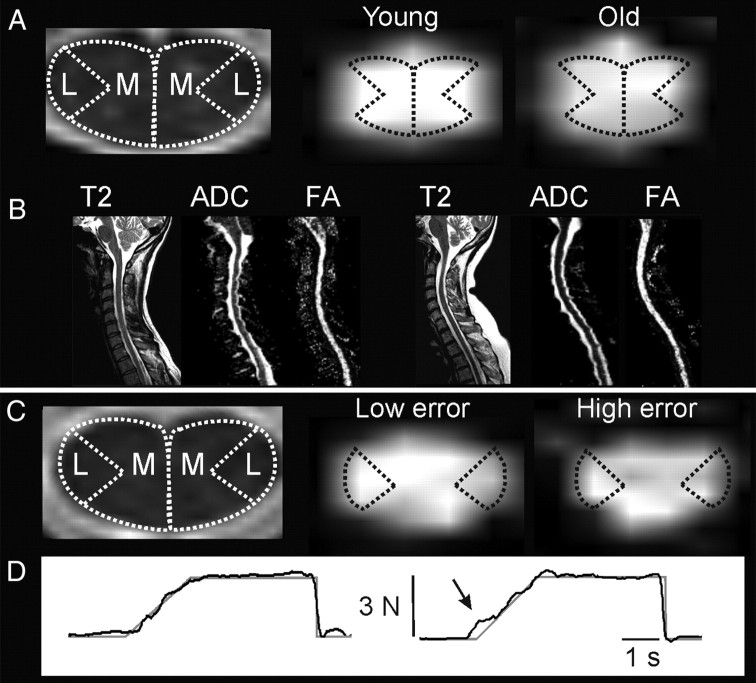
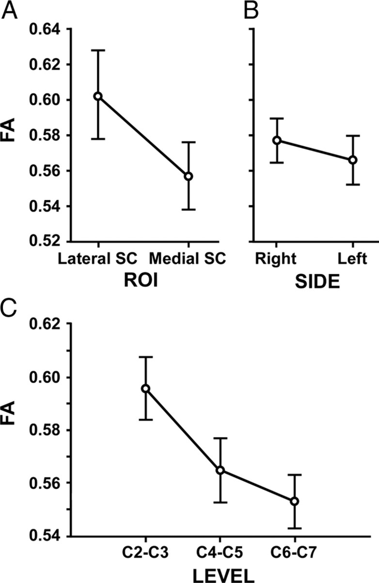
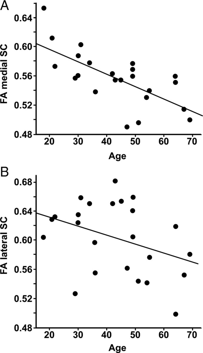
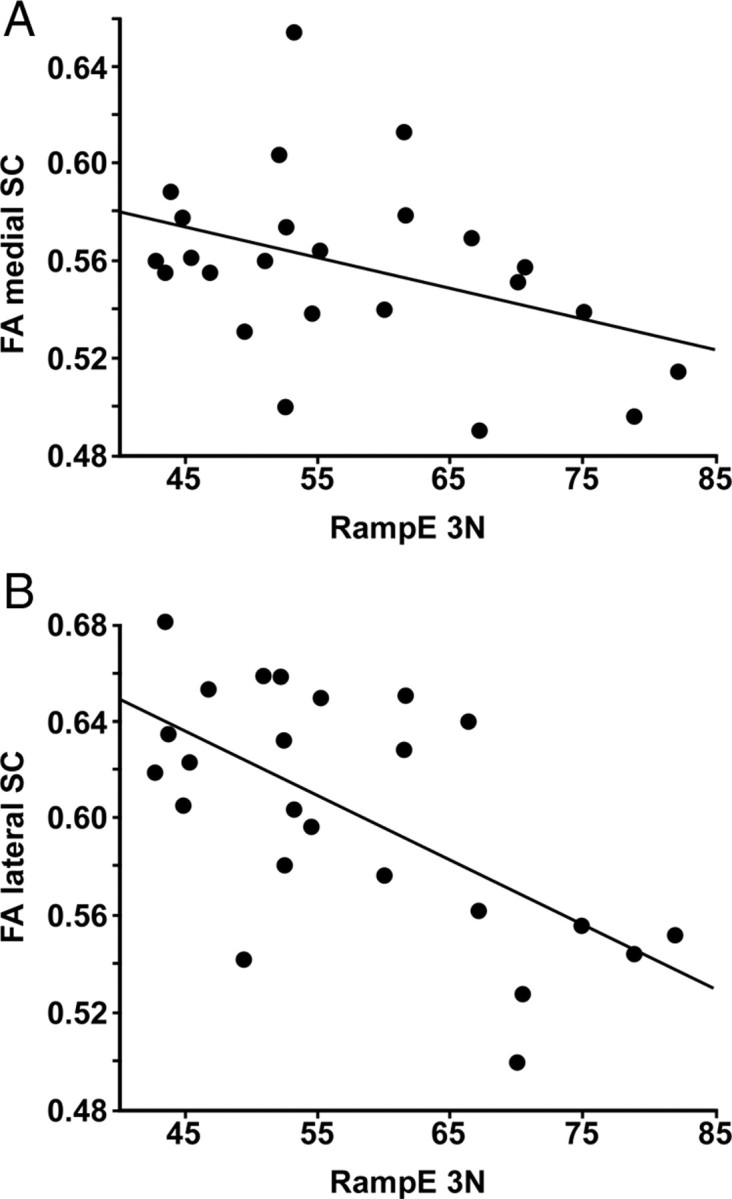
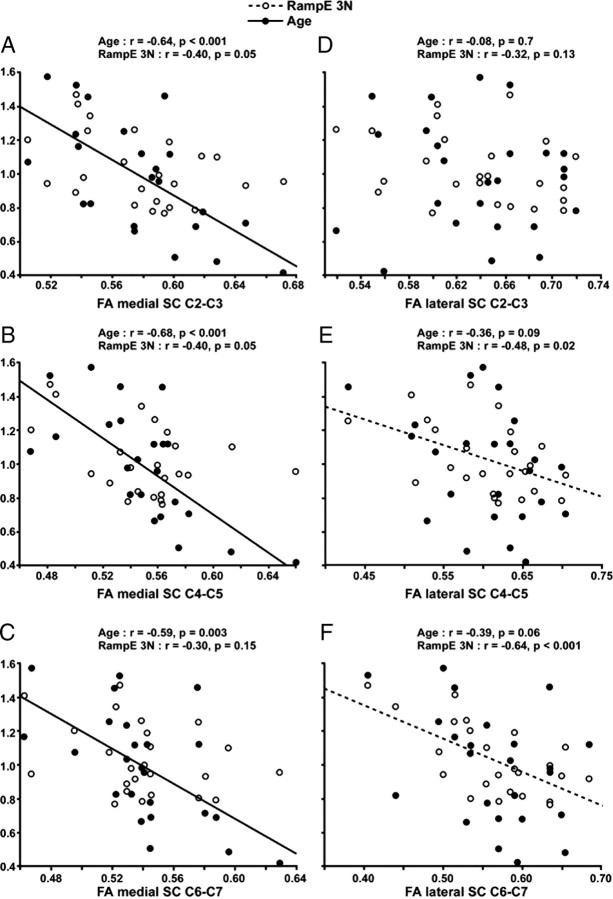
Diffusion tensor imaging (DTI) can be used to elucidate relations between CNS structure and function. We hypothesized that the degree of spinal white matter organization relates to the accuracy of control of grip force. Healthy subjects of different age were studied using DTI and visuomotor tracking of precision grip force. The latter is a prime component of manual dexterity. A regional analysis of spinal white matter [fractional anisotropy (FA)] across multiple cervical levels (C2-C3, C4-C5, and C6-C7) and in different regions of interest (left and right lateral or medial spinal cord) was performed. FA was highest at the C2-C3 level, higher on the right than the left side, and higher in the lateral than in the medial spinal cord (p < 0.001). FA of whole cervical spinal cord (C2-C7) was lower in subjects with high tracking error (r = -0.56, p = 0.004) and decreased with age (r = -0.63, p = 0.001). A multiple regression analysis revealed an independent contribution of each predictor (semipartial correlations: age, r = -0.55, p < 0.001; tracking error, r = -0.49, p = 0.003). The closest relation between FA and tracking error was found at the C6-C7 level in the lateral spinal cord, in which the corticospinal tract innervates spinal circuitry controlling hand and digit muscles. FA of the medial spinal cord correlated consistently with age across all cervical levels, whereas FA of the lateral spinal cord did not. The results suggest (1) a functionally relevant specialization of lateral spinal cord white matter and (2) an increased sensitivity to age-related decline in medial spinal cord white matter in healthy subjects.
Figures
Similar articles
-
Correlation of force control with regional spinal DTI in patients with cervical spondylosis without signs of spinal cord injury on conventional MRI.Eur Radiol. 2016 Mar;26(3):733-42. doi: 10.1007/s00330-015-3876-z. Epub 2015 Jun 27. Eur Radiol. 2016. PMID: 26123409
-
Diffusion tensor imaging of the cervical spinal cord in healthy adult population: normative values and measurement reproducibility at 3T MRI.Acta Radiol. 2014 May;55(4):478-85. doi: 10.1177/0284185113499752. Epub 2013 Aug 22. Acta Radiol. 2014. PMID: 23969263
-
Measures of spinal canal stenosis and relationship to spinal cord structure in patients with cervical spondylosis.J Neuroradiol. 2012 Oct;39(4):236-42. doi: 10.1016/j.neurad.2011.09.004. Epub 2011 Oct 26. J Neuroradiol. 2012. PMID: 22033418
-
Region-specific impairment of the cervical spinal cord (SC) in amyotrophic lateral sclerosis: A preliminary study using SC templates and quantitative MRI (diffusion tensor imaging/inhomogeneous magnetization transfer).NMR Biomed. 2017 Dec;30(12). doi: 10.1002/nbm.3801. Epub 2017 Sep 19. NMR Biomed. 2017. PMID: 28926131
-
Age-related changes of the diffusion tensor imaging parameters of the normal cervical spinal cord.Eur J Radiol. 2014 Dec;83(12):2196-2202. doi: 10.1016/j.ejrad.2014.09.010. Epub 2014 Sep 28. Eur J Radiol. 2014. PMID: 25287960
Cited by
-
Age-related weakness of proximal muscle studied with motor cortical mapping: a TMS study.PLoS One. 2014 Feb 21;9(2):e89371. doi: 10.1371/journal.pone.0089371. eCollection 2014. PLoS One. 2014. PMID: 24586726 Free PMC article.
-
Tract-specific analysis of diffusion MRI at 3T detects cervical spinal cord aberrations in multiple sclerosis.Imaging Neurosci (Camb). 2025 Jul 7;3:IMAG.a.72. doi: 10.1162/IMAG.a.72. eCollection 2025. Imaging Neurosci (Camb). 2025. PMID: 40800899 Free PMC article.
-
Quantitative assessment of column-specific degeneration in cervical spondylotic myelopathy based on diffusion tensor tractography.Eur Spine J. 2015 Jan;24(1):41-7. doi: 10.1007/s00586-014-3522-5. Epub 2014 Aug 24. Eur Spine J. 2015. PMID: 25150714
-
An Attempt of Early Detection of Poor Outcome after Whiplash.Front Neurol. 2016 Oct 20;7:177. doi: 10.3389/fneur.2016.00177. eCollection 2016. Front Neurol. 2016. PMID: 27812348 Free PMC article.
-
Manual Dexterity and Aging: A Pilot Study Disentangling Sensorimotor From Cognitive Decline.Front Neurol. 2018 Oct 29;9:910. doi: 10.3389/fneur.2018.00910. eCollection 2018. Front Neurol. 2018. PMID: 30420830 Free PMC article.
References
-
- Agosta F, Laganà M, Valsasina P, Sala S, Dall'Occhio L, Sormani MP, Judica E, Filippi M. Evidence for cervical cord tissue disorganisation with aging by diffusion tensor MRI. Neuroimage. 2007;36:728–735. - PubMed
-
- Ayabe S, Goto N, Atsumi T, Goto J, Suzuki J. Morphometric evaluation of posterior funiculus nerve fibers in relation to aging. Okajimas Folia Anat Jpn. 2005;82:35–38. - PubMed
-
- Basser PJ, Jones DK. Diffusion-tensor MRI: theory, experimental design and data analysis—a technical review. NMR Biomed. 2002;15:456–467. - PubMed
-
- Basser PJ, Pierpaoli C. Microstructural and physiological features of tissues elucidated by quantitative-diffusion-tensor MRI. J Magn Reson B. 1996;111:209–219. - PubMed
-
- Beaulieu C. The basis of anisotropic water diffusion in the nervous system: a technical review. NMR Biomed. 2002;15:435–455. - PubMed
Publication types
MeSH terms
LinkOut - more resources
Full Text Sources
Medical
Miscellaneous