Comparative Study
. 2010 Mar 17;30(11):4110-9.
doi: 10.1523/JNEUROSCI.4364-09.2010.

Temporal requirement of the protein tyrosine phosphatase Shp2 in establishing the neuronal fate in early retinal development

Affiliations
Comparative Study

Temporal requirement of the protein tyrosine phosphatase Shp2 in establishing the neuronal fate in early retinal development

Zhigang Cai et al. J Neurosci. .

Abstract

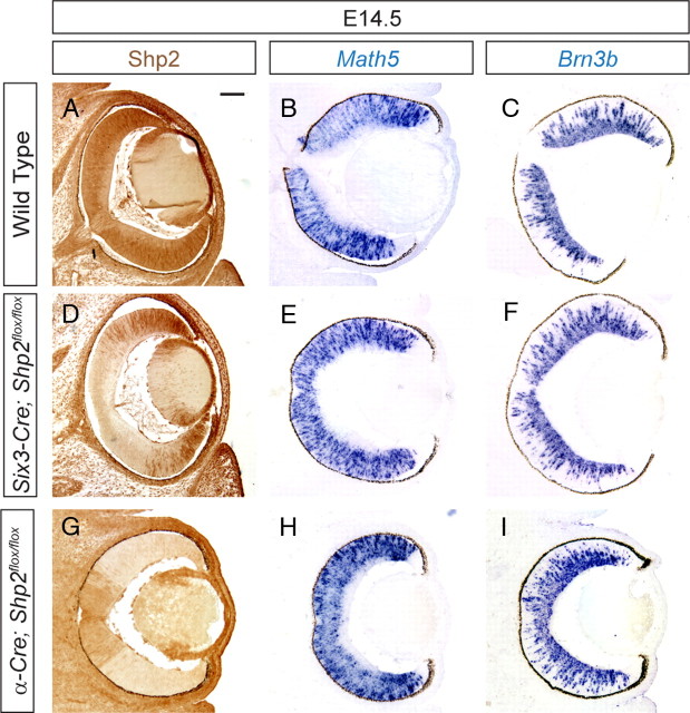
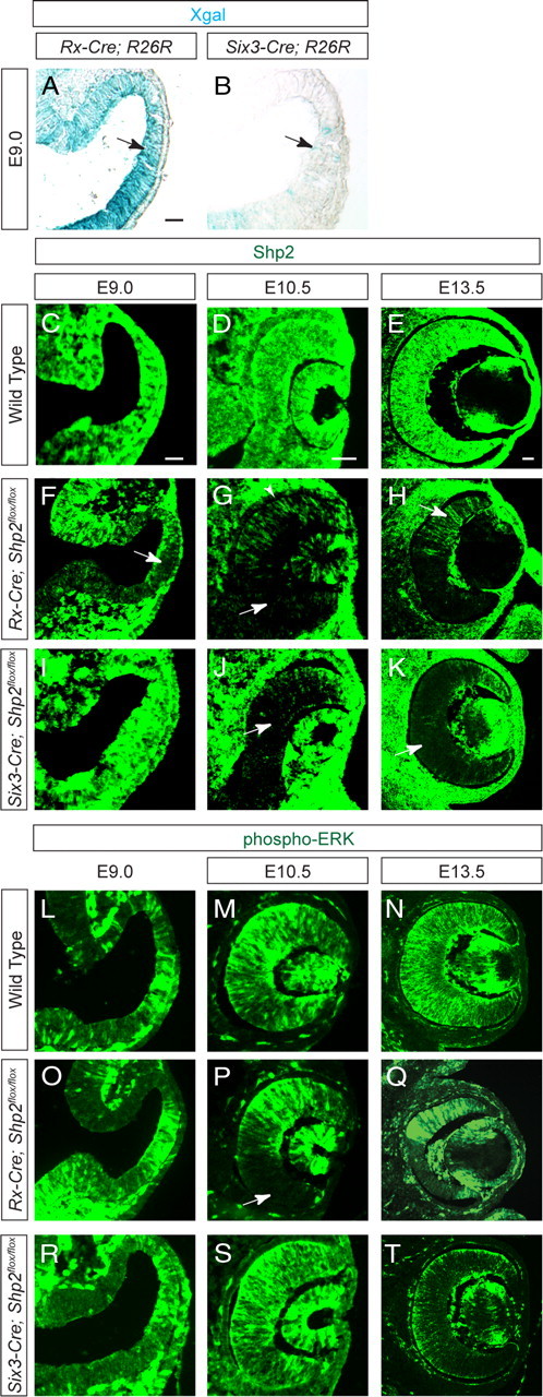
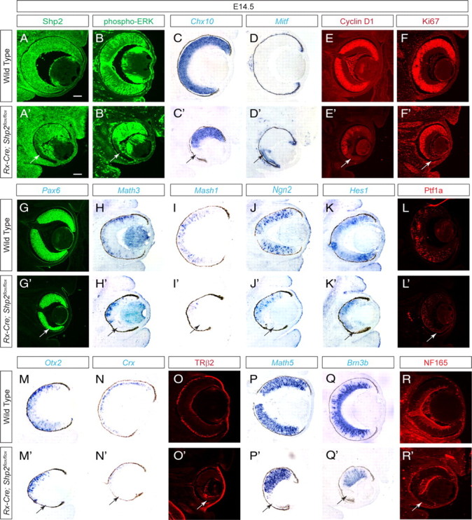
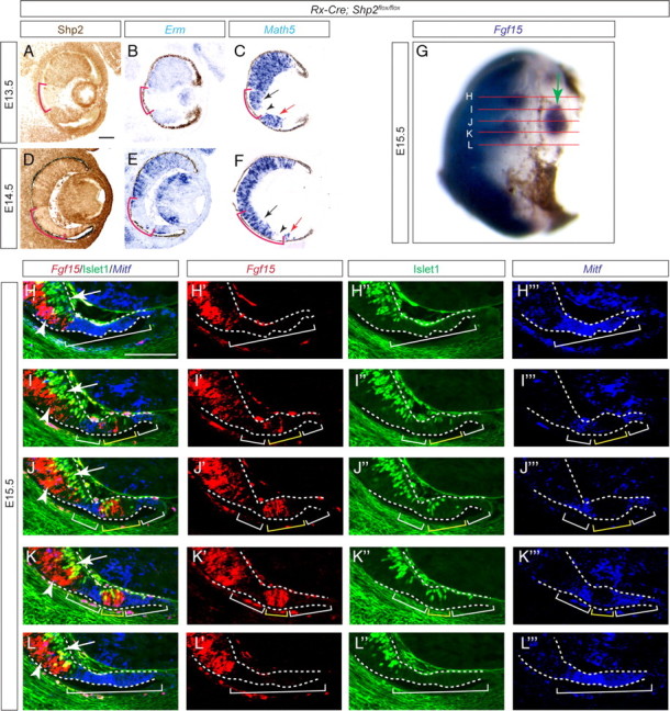
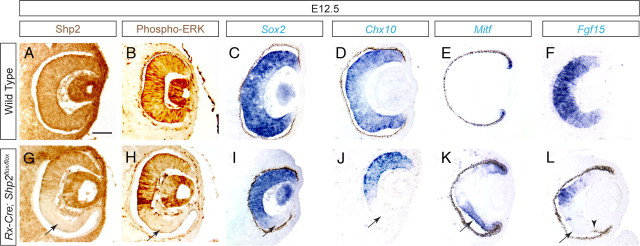
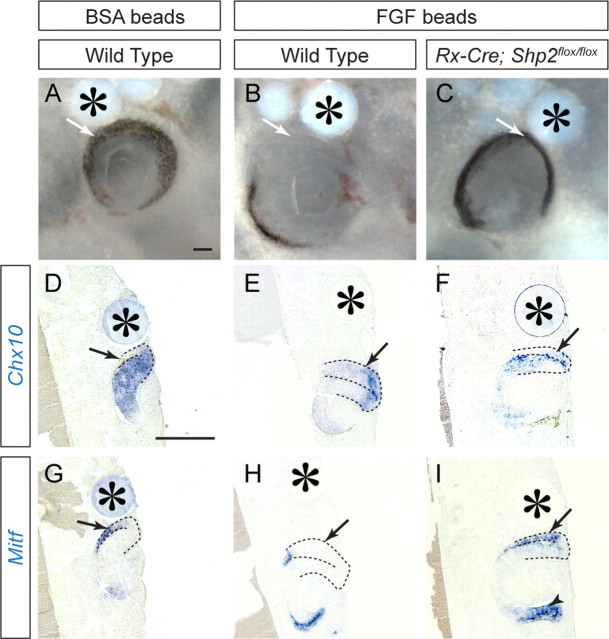
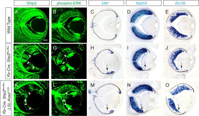
FGF signaling is critical in the development of the vertebrate retina, which differentiates in a wave-like pattern similar to that found in the Drosophila eye. In this study, we investigated the mechanism of FGF signaling in vertebrate eye development by identifying Shp2, a protein tyrosine phosphatase, as a novel factor in orchestrating retinal morphogenesis. Using a series of Shp2 conditional mutants, we have shown that Shp2 is specifically required for the initiation of retinal neurogenesis but not for the maintenance of the retinal differentiation program. By mosaic deletion of Shp2, we further demonstrated that Shp2 ablation did not prevent the spreading of the retinal differentiation wave. Shp2 instead controls the patterning of the optic vesicle by regulating the retinal progenitor factors and cell proliferation. In ex vivo culture models and genetic rescue experiments, we showed that Shp2 acts downstream to FGF signaling in retinal development and that it can be functionally substituted by activated Ras signaling. Together, these results demonstrate that Shp2 mediates FGF-Ras signaling to control retinal progenitor cell fate.


Figures

Figure 1.
Figure 1.
Lack of retinal defects in the Six3–Cre;Shp2flox/flox and α-Cre;Shp2flox/flox mutants. A–C, Expression of Shp2 and retinal differentiation genes Math5 and Brn3b in wild-type retina at E14.5. D–F, Shp2 was abolished in the center of Six3–Cre;Shp2flox/flox retina, but Math5 and Brn3b expressions remained unaffected. G–I, Shp2 was efficiently depleted in the α-Cre;Shp2flox/flox peripheral retina, but no changes were observed in Math5 and Brn3b expressions. Scale bar, 100 μm.
Figure 2.
Figure 2.
The time course of Rx–Cre- and Six3–Cre- mediated Shp2 and phospho-ERK depletion. A, B, Rx–Cre but not Six3–Cre activated strong R26R reporter expression in E9.0 optic vesicle. C–K, Reduction of Shp2 staining was first observed in the Rx–Cre;Shp2flox/flox mutant optic vesicle at E9.0 and in the Six3–Cre;Shp2flox/flox mutant at E10.5. In the Rx–Cre;Shp2flox/flox mutant, notice the loss of Shp2 staining in the future RPE (arrowhead in G) and mosaic pattern depletion of Shp2 in retina (arrow in H). L–T, As a consequence, disruption of phospho-ERK staining was observed in the Rx–Cre;Shp2flox/flox mutant optic vesicle at E10.5 but significantly delayed in Six3–Cre;Shp2flox/flox mutant retina until E13.5. The embryos at the same stages share the scale bars (50 μm). All sections are coronal with the dorsal retina at the top. Xgal, 5-Bromo-4-chloro-3-indolyl-β-d-galactopyranoside.
Figure 3.
Figure 3.
Rx–Cre-mediated Shp2 ablation disrupted retinal development. A–F, A′–F′, At E14.5, mosaic depletion of Shp2 in the Rx–Cre;Shp2flox/flox mutant retinae not only disrupted phospho-ERK staining but also resulted in the loss of Cyclin D1, Ki67, and Chx10 expression. In contrast, Mitf expression was upregulated (arrows). G–K, G′–K′, The eye field specification gene Pax6 is unchanged, but the retinal differentiation genes Math3, Math1, Ngn2, and Hes1 are all lost in the Rx–Cre;Shp2flox/flox mutant retinae (arrows). L–R, L′–R′, Loss of Ptf1a indicated the disruption of retinal amacrine and horizontal cell differentiation. Loss of Otx2, Crx, and TRβ2 indicated photoreceptor cell defects. Loss of Math5, Brn3b, and NF165 indicated retinal ganglion cells defects. Scale bars, 100 μm.
Figure 4.
Figure 4.
Shp2 ablation did not affect the neurogenic wave progression. A–F, On adjacent sections, although the Shp2 depletion pattern region exactly matched the Erm-deficient zone (red brackets), only the distal portion of the Shp2-negative zone lost Math5 expression (black arrow and arrowheads). Moreover, the wild-type cells distal to the Shp2-deficient zone still expressed Math5 (red arrows). G, Whole-mount in situ hybridization identified an island of Fgf15-expressing retinal progenitor cells isolated from proximal retina (arrow). The red lines indicated the serial sections used in H–L to fully map the Fgf15-positive domains. H–L, By combining fluorescent RNA in situ hybridization (Fgf15 and Mitf) and immunofluorescence (Islet1), we identified the Fgf15/Islet1-positive cell clusters (arrows and arrowheads within the yellow brackets) enclosed in the Mitf-positive domain (white brackets), demonstrating that neural differentiation can occur in the isolated wild-type cells in distal retina without direct contact with the proximal retina. Scale bars, 100 μm.
Figure 5.
Figure 5.
Shp2 signaling promotes the neural retinal fate against retinal pigmented epithelium fate. At E12.5, the Shp2 null cells in Rx–Cre;Shp2flox/flox mutants maintained the expression of the eye field specification gene Pax6 but lost the retinal progenitor markers Sox2 and Chx10 (A–D, G–J, arrows). There was a corresponding invasion of Mitf expression into the presumptive neural retinal domain and loss of Fgf15 expression (F, L, arrow). Notice the presence of pigmented RPE cells in the tip of the mutant retina (L, arrowhead). Scale bar, 100 μm.
Figure 6.
Figure 6.
FGF signaling depended on Shp2 to induce neural retina in optic vesicle. A, D, G, Wild-type retinal culture developed with proper demarcation of Chx10 and Mitf expressions in the presence of BSA beads (arrows). B, E, H, In wild-type cultures, FGF2 beads transformed the Mitf-expressing RPE into Chx10-expressing the neural retina (arrows). C, F, I, In Rx–Cre;Shp2flox/flox mutants, FGF2 beads were unable to induce RPE into the neural retina (arrowheads). Asterisks denote implanted beads. Scale bars, 200 μm.
Figure 7.
Figure 7.
Activated Ras signaling rescued Shp2 mutant retinal development. A–E, In the E14.5 wild-type embryos, Shp2, phospho-ERK, Math5, and Brn3b were expressed throughout the retina, whereas Mitf expression was restricted to the tip of the retina. F–J, Mosaic depletion of Shp2 in the Rx–Cre;Shp2flox/flox mutants led to a loss of phospho-ERK, Math5, and Brn3b expressions, whereas Mitf expression extended into the neural retinal region (arrows). K–O, In the Rx–Cre;Shp2flox/flox;LSL–KrasG12D compound mutants, phospho-ERK, Math5, and Brn3b expression was restored in the Shp2-negative cells, whereas Mitf was properly expressed in the tip of the retina (arrowheads). Scale bar, 100 μm.

References

    1. Brown NL, Patel S, Brzezinski J, Glaser T. Math5 is required for retinal ganglion cell and optic nerve formation. Development. 2001;128:2497–2508. - PMC - PubMed
    1. Burmeister M, Novak J, Liang MY, Basu S, Ploder L, Hawes NL, Vidgen D, Hoover F, Goldman D, Kalnins VI, Roderick TH, Taylor BA, Hankin MH, McInnes RR. Ocular retardation mouse caused by Chx10 homeobox null allele: impaired retinal progenitor proliferation and bipolar cell differentiation. Nat Genet. 1996;12:376–384. - PubMed
    1. Chen S, Wang QL, Nie Z, Sun H, Lennon G, Copeland NG, Gilbert DJ, Jenkins NA, Zack DJ. Crx, a novel Otx-like paired-homeodomain protein, binds to and transactivates photoreceptor cell-specific genes. Neuron. 1997;19:1017–1030. - PubMed
    1. Dono R, Texido G, Dussel R, Ehmke H, Zeller R. Impaired cerebral cortex development and blood pressure regulation in FGF-2-deficient mice. EMBO J. 1998;17:4213–4225. - PMC - PubMed
    1. Elshatory Y, Deng M, Xie X, Gan L. Expression of the LIM-homeodomain protein Isl1 in the developing and mature mouse retina. J Comp Neurol. 2007;503:182–197. - PMC - PubMed

Publication types

MeSH terms

Substances

LinkOut - more resources