Comparative Study
. 2010 Mar 17;30(11):4160-70.
doi: 10.1523/JNEUROSCI.3537-09.2010.

Dendritically localized transcripts are sorted into distinct ribonucleoprotein particles that display fast directional motility along dendrites of hippocampal neurons

Affiliations
Comparative Study

Dendritically localized transcripts are sorted into distinct ribonucleoprotein particles that display fast directional motility along dendrites of hippocampal neurons

Fabian Tübing et al. J Neurosci. .

Abstract

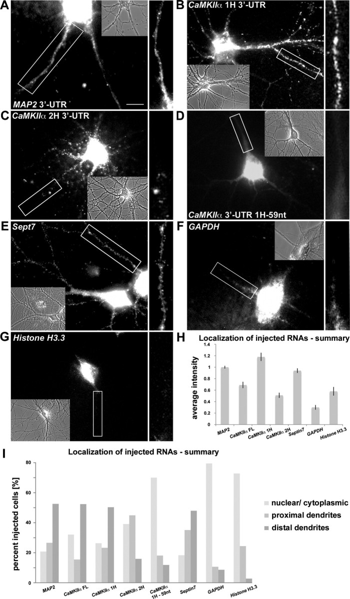
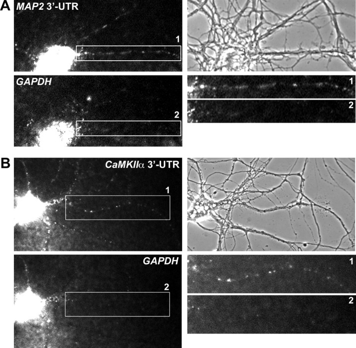
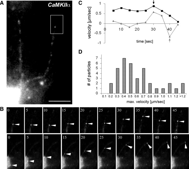
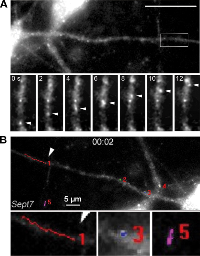
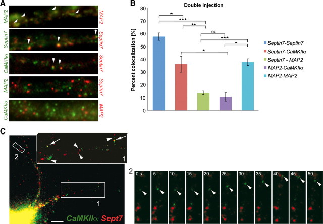
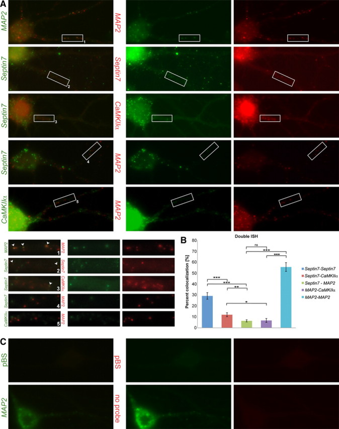
Localization of mRNAs to postsynaptic sites and their subsequent translation is thought to contribute to synapse-specific plasticity. However, the direct visualization of dendritic RNA transport in living neurons remains a major challenge. Here, we analyze the transport of Alexa-labeled RNAs microinjected into mature hippocampal neurons. We show that microinjected MAP2 and CaMKIIalpha RNAs form particles that localize into dendrites as their endogenous counterparts. In contrast, nonlocalizing RNAs or truncated CaMKIIalpha, lacking the dendritic targeting element, remain in the cell body. Furthermore, our microinjection approach allowed us to identify a novel dendritically localized RNA, Septin7. Time-lapse videomicroscopy of neurons injected with CaMKIIalpha and Septin7 RNAs demonstrates fast directional movement along the dendrites of hippocampal neurons, with similar kinetics to Staufen1 ribonucleoprotein particles (RNPs). Coinjection and simultaneous visualization of two RNAs, as well as double detection of the corresponding endogenous RNAs, reveal that neuronal transcripts are differentially sorted in dendritic RNPs.


Figures

Figure 1.
Figure 1.
Microinjected RNA forms particles that are sorted to their expected intracellular destination in cultured mature hippocampal neurons. Fifteen DIV neurons were microinjected with the following Alexa-labeled RNAs: MAP2 3′-UTR (A); CaMKIIα 3′-UTR first half (1H) (also lacking the first 31 nt of the 3′-UTR) (B); CaMKIIα 3′-UTR second half (2H) (C); CaMKIIα 3′-UTR first half lacking 59 nt (1H-59 nt) (D); Septin7 (E); GAPDH (F); and Histone H3.3 (G). Particles were imaged 5–10 min after microinjection. Phase contrast images of the respective injected neurons are shown. Insets are magnifications of the indicated dendrites on the right. H, I, Quantification of all microinjection experiments. H, Average intensity of fluorescent signal in distal dendrites. The values are normalized to the signal of microinjected MAP2 RNA. Error bars represent the SEM. I, Evaluation of the localization pattern of microinjected RNAs (>30 cells rated per RNA). Four people evaluated the data independently, in a blind manner. “Distal” reflects fluorescent signals that were found at least two cell body diameters (>25 μm) from the soma. Scale bar, 10 μm.
Figure 2.
Figure 2.
Coinjected GAPDH RNA and MAP2 or CaMKIIα RNA are sorted to their expected destinations in cultured hippocampal neurons. Coinjection of Alexa 488-labeled MAP2 3′-UTR RNA and Alexa 546-labeled GAPDH RNA (A) or Alexa 488-labeled CaMKIIα 3′-UTR and Alexa 546-labeled GAPDH RNA (B), respectively, results in dendritic localization of either MAP2 RNA or CaMKIIα 3′-UTR, whereas GAPDH RNA remains in the cell soma. Magnifications of the boxed dendritic regions and phase contrast images of the respective injected neurons are shown on the right. The size of the cell body of a typical pyramidal neuron is 10–15 μm.
Figure 3.
Figure 3.
CaMKIIα RNA particles move bidirectionally into dendrites of hippocampal neurons. A, A cultured neuron microinjected with Alexa 488-labeled CaMKIIα 3′-UTR. B, Time series of individual particles. The top panel shows bidirectional transport of a CaMKIIα RNA-containing particle (arrowheads) indicated in the boxed region of A. The bottom panel shows unidirectional (anterograde) transport of a CaMKIIα particle starting in the cell body (arrowheads). Images were taken every 5 s (supplemental Movie SM1, available at www.jneurosci.org as supplemental material). C, Kinetics of the two presented CaMKIIα particles. Negative velocities correspond to retrograde movement, and positive velocities correspond to anterograde transport. The arrows indicate the respective maximum velocities. D, Histogram of the maximum velocities of a total of 41 CaMKIIα particles. Scale bar, 10 μm.
Figure 4.
Figure 4.
Analysis of Sept7 RNA kinetics. A, Sept7 RNA particles can exhibit fast directed movement into dendrites of hippocampal neurons. Dendrite of a cultured hippocampal neuron after microinjection with Alexa-labeled Sept7 RNA. Below are images from a time-lapse video showing the movement of a Sept7 RNA particle (arrowheads) in the distal dendrite (boxed). The time span between panels corresponds to 2 s (supplemental Movie SM3, available at www.jneurosci.org as supplemental material). B, Individual Sept7 particles were tracked manually. We detected directed movement along dendrites (see particle 1). The majority of dendritic Sept7 particles either appear stationary (see particles 2, 3, 4) or seem to display diffusion (see particle 5). Scale bar, 10 μm.
Figure 5.
Figure 5.
Differential sorting of microinjected dendritic transcripts in neuronal RNPs. A, Coinjection of Alexa 546- and Alexa 488-labeled MAP2 3′-UTR–MAP2 3′-UTR, Sept7–Sept7, Sept7–CaMKIIα 1H 3′-UTR, Sept7–MAP2 3′-UTR, and MAP2 3′-UTR–CaMKIIα 1H 3′-UTR pairs. B, Quantification of the double-injection results. Colocalization ratios of Sept7–Sept7, Sept7–CaMKIIα 1H 3′-UTR, Sept7–MAP2 3′-UTR, MAP2 3′-UTR–CaMKIIα 1H 3′-UTR, and MAP2 3′-UTR–MAP2 3′-UTR were 57.48 ± 2.89, 36.04 ± 6.25, 13.98 ± 1.49, 10.69 ± 3.35, and 37.63 ± 2.63%, respectively. Error bars represent the SEM of colocalization ratios in individual cells. The asterisks denote statistical significance as determined using the two-tailed Student's t test for the complete data set. *p < 0.05; **p < 0.01; ***p < 0.001. C, Coinjection of Sept7 RNA (Alexa 546) and CaMKIIα RNA (Alexa 488; the first part of the 3′-UTR, 1H) (Fig. 1) into a representative hippocampal neuron. The arrowheads indicate particles in the enlargement (top right) of box 1 that contain either Sept7 or CaMKIIα RNA; the arrows denote colocalization of Sept7 and CaMKIIα RNA in a single particle. Movement of an individual particle found in box 2 is shown in supplemental Movie SM4 (available at www.jneurosci.org as supplemental material). The time span between individual frames is 5 s.
Figure 6.
Figure 6.
Differential sorting of endogenous dendritic transcripts in neuronal RNPs. A, Double detection with Alexa 488 and Cy3 tyramide of endogenous MAP2–MAP2, Sept7–Sept7, Sept7–CaMKIIα, Sept7–MAP2, and CaMKIIα–MAP2 pairs by fluorescence ISH. B, Quantification of the double ISH results. Colocalization ratios of Sept7–Sept7, Sept7–CaMKIIα, Sept7–MAP2, MAP2–CaMKIIα, and MAP2–MAP2 were 29.09 ± 3.00, 11.96 ± 1.82, 6.37 ± 0.98, 6.74 ± 1.77, and 55.75 ± 4.02%, respectively. Error bars represent the SEM of colocalization ratios in individual cells. The asterisks denote statistical significance as determined using two-tailed Student's t test for the complete data set. C, Double ISH specificity control with pBluescript sequences used labeled with DIG and fluorescein probes (top panel). Negative control for cross-reactivity. Cell hybridized with a fluorescein probe against MAP2 RNA shows signal only using anti-fluorescein-HRP tyramide detection and not with the subsequent anti-DIG-HRP tyramide detection (bottom panel).

Similar articles

Cited by

References

    1. Ainger K, Avossa D, Morgan F, Hill SJ, Barry C, Barbarese E, Carson JH. Transport and localization of exogenous myelin basic protein mRNA microinjected into oligodendrocytes. J Cell Biol. 1993;123:431–441. - PMC - PubMed
    1. Antar LN, Afroz R, Dictenberg JB, Carroll RC, Bassell GJ. Metabotropic glutamate receptor activation regulates fragile X mental retardation protein and FMR1 mRNA localization differentially in dendrites and at synapses. J Neurosci. 2004;24:2648–2655. - PMC - PubMed
    1. Blichenberg A, Schwanke B, Rehbein M, Garner CC, Richter D, Kindler S. Identification of a cis-acting dendritic targeting element in MAP2 mRNAs. J Neurosci. 1999;19:8818–8829. - PMC - PubMed
    1. Blichenberg A, Rehbein M, Müller R, Garner CC, Richter D, Kindler S. Identification of a cis-acting dendritic targeting element in the mRNA encoding the alpha subunit of Ca2+/calmodulin-dependent protein kinase II. Eur J Neurosci. 2001;13:1881–1888. - PubMed
    1. Bramham CR, Wells DG. Dendritic mRNA: transport, translation and function. Nat Rev Neurosci. 2007;8:776–789. - PubMed

Publication types

MeSH terms

LinkOut - more resources