Comparative Study
. 2010 Apr 15;184(8):4215-27.
doi: 10.4049/jimmunol.0902995. Epub 2010 Mar 17.

Adoptive transfer of tumor-specific Tc17 effector T cells controls the growth of B16 melanoma in mice

Affiliations
Comparative Study

Adoptive transfer of tumor-specific Tc17 effector T cells controls the growth of B16 melanoma in mice

Maria de la Luz Garcia-Hernandez et al. J Immunol. .

Abstract

In vitro generated OVA-specific IL-17-producing CD8 T effector cells (Tc17) from OT-1 mice, adoptively transferred into B16-OVA tumor-bearing mice, controlled tumor growth in early and late stage melanoma. IL-17, TNF, and IFN-gamma from the Tc17 effectors all played a role in an enhanced recruitment of T cells, neutrophils, and macrophages to the tumor. In addition, Tc17 cells and recently recruited, activated neutrophils produced further chemokines, including CCL3, CCL4, CCL5, CXCL9, and CXCL10, responsible for the attraction of type 1 lymphocytes (Th1 and Tc1) and additional neutrophils. Neutrophils were rapidly attracted to the tumor site by an IL-17 dependent mechanism, but at later stages the induction of the chemokine CXCL2 by Tc17-derived TNF and IFN-gamma contributed to sustain neutrophil recruitment. Approximately 10-50 times as many Tc17 effectors were required compared with Tc1 effectors to exert the same level of control over tumor growth. The recruitment of neutrophils was more prominent when Tc17 rather than Tc1 were used to control tumor and depletion of neutrophils resulted in a diminished capacity to control tumor growth.


Conflict of interest statement

Disclosures

The authors have no conflicting financial interests.

Figures
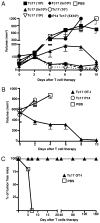
FIGURE 1
FIGURE 1
A, Tc17 cells control the growth of established melanoma tumor. Seven groups of C57BL/6 mice were injected with 2 × 10⁵ OVA-expressing B16 cells at day −7. At day 0 five groups of mice were treated with different numbers of Tc17 generated from OT-1 mice. As controls, the other two groups were treated with Tc17 cell generated from P14 mice (closed circles) or PBS (open squares). Tumor growth was measured three times a week using an engineer caliper. Results represent the mean ± SE of five mice from three separate experiments with similar results. B, Tc17 cells control growth of large tumors. The 5 × 10⁶ Tc17 OT-1 (closed triangles) cells were adoptively transferred at day 0 into 12 d tumor-bearing C57BL/6 mice. As controls, two groups of mice were treated with LCMV-specific Tc17 (open circles) or with PBS (open squares). Tumor growth was measured three times at week using an engineer caliper. Results represent the mean ± SE of five mice. C, Tc17 cells prevent tumor implantation and induce long-lasting tumor immunity. The 5 × 10⁶ Tc17 (closed triangles) cells were adoptively transferred at day −1 into C57BL/6 mice, whereas control mice received PBS (open squares). At day 0 mice were challenged with 2 × 10⁵ B16-OVA cells and tumor growth was monitored. Mice that did not develop tumor were rechallenged twice (day 45 and 120). Mean ± SE, five mice.
FIGURE 2
FIGURE 2
A, IFN-γ and TNF, but not perforin, are required for complete control of tumor growth. Groups of tumor-bearing mice were treated with 5 × 10⁶ Tc17 cells generated from OT-1 (closed squares), perforin- (closed triangles), IFN-γ– (close inverted triangles) and TNF- (closed diamonds) deficient OT-1 mice. A control group was treated with LCMV-specific P14 Tc17 (open circles). Tumor growth was monitored three times a week. Data represent the mean ±SE of 10 mice in two separate experiments. OT-1 Tc17 versus OT-1.IFN-γ knockout (KO) Tc17 p = 0.006, two tailed. OT-1 Tc17 versus OT-1.TNF KO Tc17 p = 0.028, two tailed. OT-1 Tc17 versus P14 Tc17 p = 0.005, two tailed. B and C, Control of tumor growth by Tc17 is associated to production of TNF and IFN-γ. C57BL/6-, IFN-γ−, and TNF-deficient mice were injected with 2 × 10⁵ B16-OVA cells. At day 7 mice were treated with different doses of wild-type, IFN-γ– (B) or TNF- (C) deficient Tc17 in all four permutations as shown in the key. The percentage of tumor volume reduction was calculated at day 6 and plotted against number of Tc17. Results represent the mean ± SE of five mice from two separate experiments with similar results.
FIGURE 3
FIGURE 3
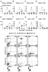
Tc17 therapy induces an increased recruitment of immune cells into tumor. Tumor-bearing mice were treated with 5 × 10⁶ Tc17 wild-type (closed triangles), IFN-γ– (closed squares), and TNF- (inverted closed triangles) deficient OT-1 cells and then, sacrificed at day 2, 4, 6, and 8 after Tc17 transfer. TILs were recovered from fresh tumors, stained with SIINFEKL-APC and specific Abs for CD8, CD45.2, CD45.1, IFN-γ, TNF, and IL-17 and analyzed by flow cytometry for the markers indicated in panels a–h. Absolute numbers of positive cells per tumor were calculated and normalized per gram of tumor. Representative plots from Thy1.2+, CD45.2+, IL-17+, IFN-γ+, or TNF+ are shown. Results represent the mean ± SE of five mice from two separate experiments with similar results.
FIGURE 4
FIGURE 4
Tc17 therapy induces recruitment of macrophages and neutrophils into tumor. A, Tumor-bearing mice were treated with 106 Tc1 or 5 × 10⁶ Tc17-, TNF KO and IFN-γ KO OT-1 cells. Control mice were treated with PBS. Mice were sacrificed at day 2, 4, 6, and 8 after Tc17 transfer. Tumor infiltrating cells, recovered from fresh tumors, were stained with specific Abs (F4/80, CD11b, CD11c, 7/4, Ly6G, and Ly6C) and analyzed by flow cytometry. Absolute numbers of neutrophils and macrophages per tumor were calculated and normalized per gram of tumor. Representative data of three independent experiments with similar results is shown. Mean of four mice; bars, SE. B, Elevated levels of mRNA for CXCL2, CCL2, and IL-17 correlated with neutrophil and macrophage recruitment. Tumors from the Tc17-treated mice and the control group were harvested and weighed. DNA-free RNA was isolated from tumors of each experimental group and reversed transcribed. Quantitative PCR was performed to determine relative changes in levels of mRNA. mRNA expression levels of each gene (panels a–e) were calculated as described in Materials and Methods. Data represent the mean ± SE mRNA level of five mice per group.
FIGURE 5
FIGURE 5
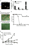
Purification and characterization of neutrophils from tumors of mice treated with Tc17. A, Mice with 7 d established tumors were treated with 5 × 10⁶ Tc17 OT-1 cells or PBS and then, tumor infiltrating neutrophils were purified at day 4. Cytospin preparations from treated and control group were stained with anti-mouse GR-1 and 7/4. B, Elevated gene expression in neutrophils from Tc17-treated mice. DNA-free RNA was isolated from purified neutrophils of each experimental group and reversed transcribed. Quantitative PCR was performed to determine relative changes in levels of mRNA for TRAIL, COX2, MPO, and iNOS (iNOS2). The relative level of mRNA expression for each gene in each group was first normalized to the expression of GAPDH and then, normalized to the level of mRNA expression in neutrophils from control group. Data represent the mean ± SE mRNA level of a pool of 15 mice per group of two separate experiments with similar results. C, Neutrophils purified from Tc17-treated mice kill tumor cells in vitro. The 2 × 10⁶ tumor infiltrating neutrophils from Tc17- (closed triangles) or PBS- (open squares) treated group were cocultured with 106 B16-OVA cells. A movie recorded events during a 2-h period using a Zeiss AxioVert 200M. Cells were imaged with differential interference contrast optics and the fluorescent signals were captured with the appropriate filter sets for GFP and propidium iodide. In stills from the movie, GFP+ neutrophils, isolated from treated mice, forming clusters with dying melanoma cells are shown in the upper panel. In contrast only a few neutrophils, isolated from tumors of control group were found in close proximity to live melanoma cells, lower panel. D, Neutrophils limit tumor growth in vivo. The 2 × 10⁵ surviving tumor cells from the previously described panel (see text for more details) were injected into C57BL/6. Tumor growth was followed and the percentage of tumor-free mice was plotted. Mean ± SE, five mice per group. E, Neutrophils play an important role in the control of tumor growth. Two groups of tumor-bearing mice were treated with Tc17 OT-1 cells at day 0 (triangles). At day −1, 0, 1, 2, 4, and 6 one group of mice received intratumor injections of mouse anti-Ly6G (250 μg/mouse, closed triangles), isotype control (open triangles), or PBS (open squares). Tumor growth was monitored three times per week. Data represent the mean ± SE of five mice in one of two separate experiments.
FIGURE 6
FIGURE 6
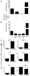
Attraction of neutrophils by Tc17 cells depends on IL-17, IFN-γ, and TNF production. A, Activated Tc17 effectors attract neutrophils. Mouse bone marrow neutrophils were placed in a transwell chamber containing 2 × 10⁶ in vitro generated wild-type Tc17 OT-1 and IFN-γ– or TNF-deficient OT-1 cells in the bottom chamber. Neutrophils that migrated toward CXCL2, wild-type, or cytokine-deficient Tc17 effectors were collected and analyzed by FACS. The results are expressed as the mean ± SEM of the CI (see Materials and Methods) of triplicate cultures. B, Neutrophils need TNFR or IFN-γ R for activation. Tc17 cells prepared from wild-type OT-1 mice were stimulated for 3 h with PMA and ionomycin and plated on the bottom of a chemotaxis chamber. Then, 106 bone marrow neutrophils from different mouse strains, C57BL/6 Thy1.1, IL-17R, TNFR, or IFN-γ R deficient were deposited on the top chamber. As a positive control for neutrophil migration some wells were filled with media containing 250 ng/ml CXCL2. The number of transmigrated neutrophils was evaluated by FACS and the CI was calculated. C, Chemokine expression of activated neutrophils. Tumor infiltrating neutrophils were purified from tumor-bearing mice treated with Tc17 OT-1 cells or PBS. Then DNA-free RNA was isolated and reversed transcribed. Quantitative PCR was performed to determine relative changes in levels of CXCL9, CXCL10, CCL3, CCL4, CCL5, and CXCL2. The relative level of mRNA expression for each gene in each group was first normalized to the expression of GAPDH and then, normalized to the level of mRNA expression in neutrophils from PBS-treated group. Data represent the mean ± SE of a pool of 15 and 30 mice from Tc17- and PBS-treated group respectively of two separate experiments with similar results.
FIGURE 7
FIGURE 7
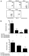
Chemokine production by Tc17 effectors. A, Tc17 cells produce chemokines involved in the attraction of Th1 cells. Four-day in vitro generated Tc17 OT-1 cells were stimulated with SIINFEKL peptide for 3 h in RPMI media containing brefeldin A. CD8 T cells were stained with Abs specific for CXCL9, CXCL10, CCL5, CCL3, and CCL4 and analyzed by FACs. B, Chemotactic activity of chemokines produced by Tc17 cells evaluated by chemotaxis assays. Tc17 cells or Tc17 and blocking Abs against the indicated chemokines were put in the bottom of a chemotaxis chamber. The 106 Th1 cells were deposited in the top chamber, and the Th1 cells that migrated to the bottom chamber were enumerated by FACS analysis after 1 h of incubation. The CI was calculated for all groups (see Materials and Methods).
FIGURE 8
FIGURE 8
Early recruitment of CXCR3+ effector cells is important for rapid induction of tumor immunity. A, At day −10 groups of C57BL/6-(triangles) or CXCR3-deficient (circles) mice were injected with 2 × 10⁵ OVA-expressing B16 cells. At day 0 two groups of mice were treated with 5 × 10⁶ Tc17 generated from OT-1 mice (closed symbols). PBS injected wild-type (open triangles) and CXCR3 deficient (open circles) served as controls. Tumor growth was measured three times at week using an engineer caliper. Results represent the mean ± SE of nine mice in two independent experiments. B, CCR5-deficient (triangles) or wild-type (circles) mice were injected with 2 × 10⁵ OVA-expressing B16 cells. At day 0 mice were treated with 5 × 10⁶ Tc17 generated from OT-1 mice (closed symbols) or PBS (open symbols). Seven days later mice were injected as indicated and tumor growth was measured three times per week. Data represent the mean ± SE of four mice in one of two experiments with similar results.
FIGURE 9
FIGURE 9
The relative effectiveness of Tc17 and Tc1 effectors in controlling tumor growth. A, Tumor growth at an intradermal site. Mice with 7 d established intradermal tumors were injected i.v. with graded numbers of Tc17 effectors, 105 Tc1, or left untreated. Tumor growth was measured three times at week using an engineer caliper. Results are representative of three experiments. B, Tumor growth as lung metastases 2 × 10⁵ B16-OVA were injected i.v. on day −7 to develop lung metastases and varying numbers of Tc17 or Tc1 effectors were injected i.v. on day 0. A control group was left untreated. Mouse survival was followed for 50 d. n = 5. The log of the number injected cells was plotted against the prolongation of survival calculated as actual time of death minus the mean time of death of the untreated group (single experiment).

Similar articles

Cited by

References

    1. Chang AE, Aruga A, Cameron MJ, Sondak VK, Normolle DP, Fox BA, Shu S. Adoptive immunotherapy with vaccine-primed lymph node cells secondarily activated with anti-CD3 and interleukin-2. J Clin Oncol. 1997;15:796–807. - PubMed
    1. Chang AE, Yoshizawa H, Sakai K, Cameron MJ, Sondak VK, Shu S. Clinical observations on adoptive immunotherapy with vaccine-primed T-lymphocytes secondarily sensitized to tumor in vitro. Cancer Res. 1993;53:1043–1050. - PubMed
    1. Yee C, Thompson JA, Byrd D, Riddell SR, Roche P, Celis E, Greenberg PD. Adoptive T cell therapy using antigen-specific CD8+ T cell clones for the treatment of patients with metastatic melanoma: in vivo persistence, migration, and antitumor effect of transferred T cells. Proc Natl Acad Sci USA. 2002;99:16168–16173. - PMC - PubMed
    1. Grimm EA, Robb RJ, Roth JA, Neckers LM, Lachman LB, Wilson DJ, Rosenberg SA. Lymphokine-activated killer cell phenomenon. III. Evidence that IL-2 is sufficient for direct activation of peripheral blood lymphocytes into lymphokine-activated killer cells. J Exp Med. 1983;158:1356–1361. - PMC - PubMed
    1. Mulé JJ, Shu S, Schwarz SL, Rosenberg SA. Adoptive immunotherapy of established pulmonary metastases with LAK cells and recombinant interleukin-2. Science. 1984;225:1487–1489. - PubMed

Publication types

MeSH terms