. 2010 Jun;298(6):R1588-96.
doi: 10.1152/ajpregu.00580.2009. Epub 2010 Mar 17.

Growth hormone rescues hippocampal synaptic function after sleep deprivation

Affiliations

Growth hormone rescues hippocampal synaptic function after sleep deprivation

Eunyoung Kim et al. Am J Physiol Regul Integr Comp Physiol. 2010 Jun.

Abstract

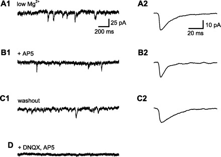
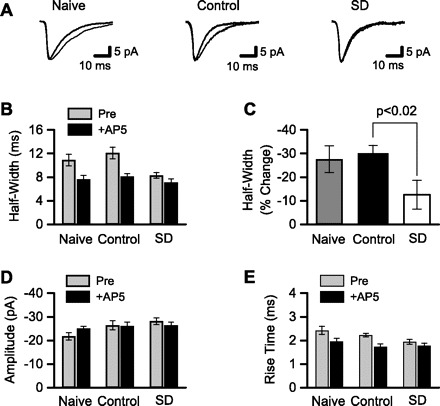
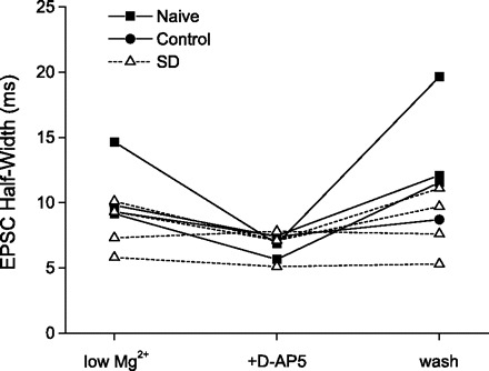
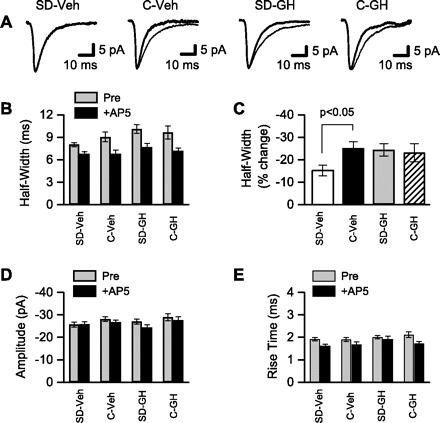
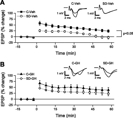
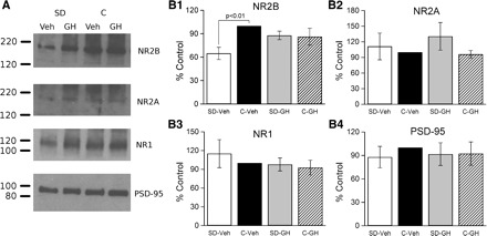
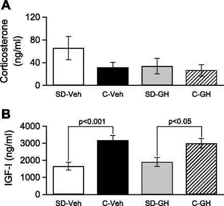
Sleep is required for, and sleep loss impairs, normal hippocampal synaptic N-methyl-D-aspartate (NMDA) glutamate receptor function and expression, hippocampal NMDA receptor-dependent synaptic plasticity, and hippocampal-dependent memory function. Although sleep is essential, the signals linking sleep to hippocampal function are not known. One potential signal is growth hormone. Growth hormone is released during sleep, and its release is suppressed during sleep deprivation. If growth hormone links sleep to hippocampal function, then restoration of growth hormone during sleep deprivation should prevent adverse consequences of sleep loss. To test this hypothesis, we examined rat hippocampus for spontaneous excitatory synaptic currents in CA1 pyramidal neurons, long-term potentiation in area CA1, and NMDA receptor subunit proteins in synaptic membranes. Three days of sleep deprivation caused a significant reduction in NMDA receptor-mediated synaptic currents compared with control treatments. When rats were injected with growth hormone once per day during sleep deprivation, the loss of NMDA receptor-mediated synaptic currents was prevented. Growth hormone injections also prevented the impairment of long-term potentiation that normally follows sleep deprivation. In addition, sleep deprivation led to a selective loss of NMDA receptor 2B (NR2B) from hippocampal synaptic membranes, but normal NR2B expression was restored by growth hormone injection. Our results identify growth hormone as a critical mediator linking sleep to normal synaptic function of the hippocampus.


Figures

Fig. 1.
Fig. 1.
Measurement of N-methyl-d-aspartate receptor (NMDAR)-dependent component of spontaneous synaptic current. All data in this figure are from a single CA1 pyramidal neuron, clamped near the normal resting membrane potential (−70 mV). Spontaneous excitatory postsynaptic currents (sEPSCs) were recorded with low extracellular Mg2+ (50 μM) to allow NMDAR current flow at the negative holding potential and with GABA receptors blocked by a combination of extracellular bicuculline (10 μM) and intracellular Cs+. A1: 2-s sample of membrane current. sEPSCs were identified using an automated method and verified by visual inspection. A2: averaged sEPSCs from a 5-min baseline period. sEPSC amplitude, rise time (10–90%), and duration at half amplitude (half-width) were measured . B1: membrane current after application of d(−)-2-amino-5-phosphonopentanoic acid (d-AP5; 50 μM) to block NMDARs. B2: averaged sEPSCs from a 5-min period after wash-in of d-AP5. Note the decrease in EPSC half-width (from 14.7 to 6.9 ms) with little change in sEPSC amplitude, consistent with the selective block of slower NDMAR-mediated synaptic currents by d-AP5. C1: washout of d-AP5 restored sEPSCs to baseline. C2: averaged sEPSC after d-AP5 washout. Half-width (19.7 ms) was similar to baseline, indicating recovery of NMDAR-mediated synaptic current. D: application of both 6,7-dinitroquinoxaline-2,3(1H, 4H)-dione (DNQX) and d-AP5 abolished all synaptic currents.
Fig. 2.
Fig. 2.
NMDAR-mediated, but not α-amino-3-hydroxy-5-methyl-4-isoxazole propionic acid receptor (AMPAR)-mediated, synaptic currents were reduced after sleep deprivation. Whole cell patch-clamp was used to record sEPSCs. A: averaged sEPSCs from 3 different CA1 pyramidal neurons. sEPSCs were recorded in low-Mg2+ ACSF with GABA receptors blocked (thin line) and after addition of the NMDAR antagonist d-AP5 (thick line). Recordings are from single neurons in slices from (left to right): naive animal housed in standard animal cage, control animal kept on large platform over water, and sleep-deprived (SD) animal kept on small platform over water. The d-AP5-sensitive portion of each sEPSC reflects the NMDAR-mediated component of synaptic current. B: NMDAR-mediated synaptic currents were quantified by measuring EPSC half-width before and after application of d-AP5. sEPSC half-widths were averaged across all cells from the same treatment condition. d-AP5 substantially reduced sEPSC half-width in cells (n = 11, 9) from naive and control animals, but not in cells (n = 9) from SD animals. C: mean change in sEPSC half-width after d-AP5 application was calculated from the data shown in B. The change in half-width was significantly smaller in the SD group compared with control. sEPSC amplitudes (D) and rise times (E) are shown. After NMDARs were blocked by d-AP5, there were no differences among the 3 groups in sEPSC half-width (B), amplitude (D), or rise time (E), indicating that there was no effect of treatment on AMPAR function.
Fig. 3.
Fig. 3.
In cells that could be held long enough for complete washout of d-AP5, NMDAR function recovered to baseline levels. sEPSC half-width is plotted for each cell that was held long enough to complete washout of d-AP5. In cells from naive and control animals, sEPSC half-widths were consistently and substantially reduced from baseline (low Mg2+) during d-AP5 application as shown earlier (see Fig. 2), but fully recovered following washout of d-AP5 (wash). Even in this reduced sample size, it is evident that sEPSC half-widths in cells from SD animals were less sensitive to d-AP5 application, as shown above in Fig. 2; however, when half-width was reduced by d-AP5 in these cells, it recovered completely after washout.
Fig. 4.
Fig. 4.
NMDAR-mediated synaptic currents were reduced after sleep deprivation, but were restored by growth hormone (GH) treatment. Whole cell patch-clamp was used to record sEPSCs. A: averaged sEPSCs from 4 different CA1 pyramidal neurons. sEPSCs were recorded in low-Mg2+ ACSF with GABA receptors blocked (thin line) and after addition of the NMDAR antagonist, d-AP5 (thick line). Recordings are from single neurons in slices from (left to right): SD animals that received daily injections of saline vehicle (SD-Veh), control animals that received daily injections of Veh (C-Veh), SD animals that received daily injections of GH (SD-GH), and control animals that received daily injections of GH (C-GH). The d-AP5-sensitive portion of each sEPSC reflects the NMDAR-mediated component of synaptic current. B: NMDAR-mediated synaptic currents were quantified by measuring sEPSC half-width before and after application of d-AP5. sEPSC half-widths were averaged across all cells from the same treatment condition. sEPSC half-width was minimally affected by d-AP5 in cells (n = 13) from SD-Veh animals, but was substantially reduced in cells (n = 13, 15, 11) from the remaining 3 treatment conditions. C: mean change in sEPSC half-width after d-AP5 application was calculated from the data shown in B. The change in half-width was significantly smaller in the SD-Veh group compared with C-Veh. In contrast, sEPSC half-width was restored to control levels in SD-GH animals. sEPSC amplitudes (D) and rise times (E) are shown. Following block of NMDARs with d-AP5, there were no differences among the 4 groups in sEPSC half-width (B), amplitude (D), or rise time (E), indicating no effect of sleep deprivation or GH on AMPAR function.
Fig. 5.
Fig. 5.
Long-term potentiation (LTP) was significantly impaired after sleep deprivation, but was rescued by GH treatment. For each slice, field excitatory postsynaptic potential (EPSP) slopes were normalized relative to baseline, averaged over consecutive 5-min periods, and then averaged across all slices in the same group. Theta burst stimulation (TBS; at time = 0 min) was used to test for LTP. A: LTP was greatly reduced in slices from the SD-Veh group compared with slices from C-Veh animals. By 45 min following TBS, LTP in the SD-Veh group had completely decayed, but LTP remained throughout the 60-min post-TBS recording period in slices from the C-Veh group. At 50–60 min post-TBS, the change in EPSPs was significantly greater in the C-Veh group compared with the SD-Veh group. Insets show averaged EPSPs from representative slices in C-Veh and SD-Veh groups obtained over the final 5 min of the baseline period and the final 5 min of the recording. B: GH treatment restored LTP in slices from SD animals to control levels. LTP was maintained at equivalent levels throughout the 60-min post-TBS recording in both SD-GH and C-GH slices. At 50–60 min post-TBS, there was no significant difference between the 2 groups. Insets show averaged EPSPs from representative slices in C-GH and SD-GH groups obtained over the final 5 min of the baseline period and the final 5 min of the recording.
Fig. 6.
Fig. 6.
Hippocampal synaptic NMDAR subunit 2B (NR2B) expression was decreased after sleep deprivation but restored by GH injection. Proteins were isolated from synaptosomal membranes, separated by gel electrophoresis, transferred to nitrocellulose membranes, and probed using antibodies specific for NMDAR subunits (NR2B, NR2A, NR1) and the postsynaptic density protein-95 (PSD-95). A: results from 1 replication of this experiment. Each replication included 4 animals, 1 from each group (SD-Veh, SD-GH, C-Veh, C-GH). In this replication, NR2B expression (top) was substantially reduced in SD-Veh animals, but was restored to control level by GH injection, whereas NR2A and NR1 subunits (middle rows) were less affected. Subunit expression did not differ between control groups (GH or Veh). There were no differences in PSD-95. B, 1–4: this experiment was repeated a total of 5 times (a total of 5 animals/treatment condition). Protein expression was quantified by film densitometry and normalized within each blot to C-Veh. Normalized values were averaged across blots (animals). The only consistent change in protein expression was for the NR2B subunit (B1), which was significantly reduced in the SD-Veh group compared with the C-Veh group, and that was restored by GH injection (no significant difference between SD-GH and C-GH). B, 2–4: there were no differences between the SD-Veh and C-Veh or SD-GH and C-GH groups for NR2A, NR1, and PSD-95.
Fig. 7.
Fig. 7.
Serum corticosterone was not altered by SD or GH treatment; however, serum IGF-I was reduced by sleep deprivation but was not restored by GH treatment. Serum hormone concentrations were determined by ELISA. A: serum corticosterone concentration was not significantly different among the 4 groups examined. B: serum IGF-I concentration was significantly lower in SD-Veh animals compared with C-Veh animals. A significant difference remained between SD-GH and C-GH animals, indicating that GH treatment did not restore serum IGF-I following SD.

Similar articles

Cited by

References

    1. Arwert LI, Deijen JB, Müller M, Drent ML. Long-term growth hormone treatment preserves GH-induced memory and mood improvements: a 10-year follow-up study in GH-deficient adult men. Horm Behav 47: 343–349, 2005 - PubMed
    1. Barnes CA. Long-term potentiation and the ageing brain. Philos Trans R Soc Lond B Biol Sci 358: 765–772, 2003 - PMC - PubMed
    1. Barria A, Malinow R. NMDA receptor subunit composition controls synaptic plasticity by regulating binding to CaMKII. Neuron 48: 289–301, 2005 - PubMed
    1. Bliss TV, Collingridge GL. A synaptic model of memory: long-term potentiation in the hippocampus. Nature 361: 31–39, 1993 - PubMed
    1. Brandenberger G, Gronfier C, Chapotot F, Simon C, Piquard F. Effect of sleep deprivation on overall 24 h growth-hormone secretion. Lancet 356: 1408, 2000 - PubMed

Publication types

LinkOut - more resources