. 2010 May 27;115(21):4273-83.
doi: 10.1182/blood-2009-09-241356. Epub 2010 Mar 17.

Cystine 186-cystine 209 disulfide bond is not essential for the procoagulant activity of tissue factor or for its de-encryption

Affiliations

Cystine 186-cystine 209 disulfide bond is not essential for the procoagulant activity of tissue factor or for its de-encryption

Hema Kothari et al. Blood. .

Abstract

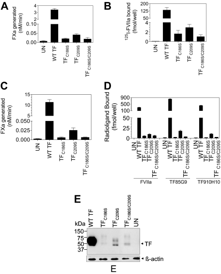
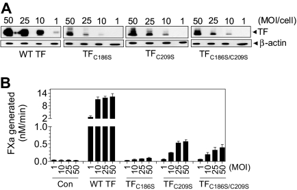
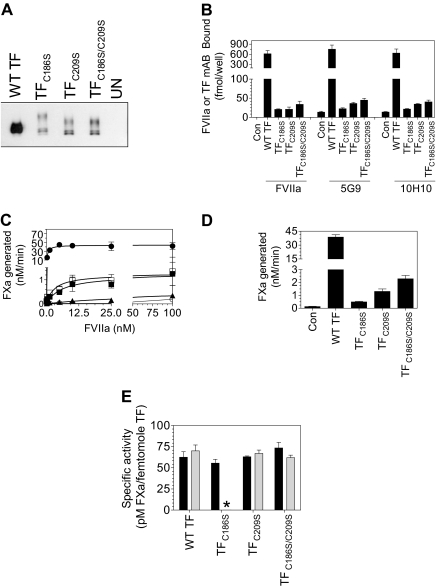
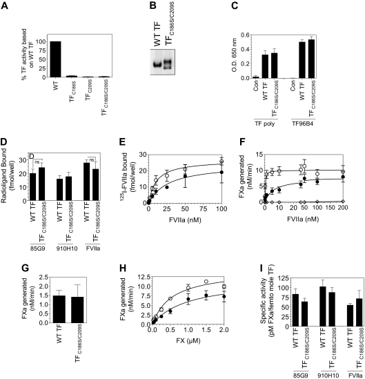
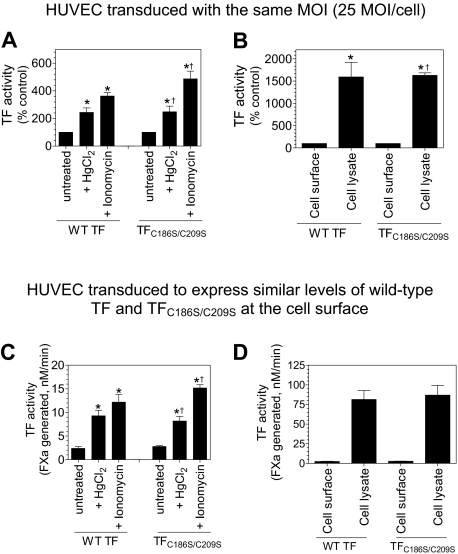
Tissue factor (TF) on cell surfaces resides mostly in a cryptic state. It is not entirely clear how cryptic TF differs from procoagulantly active TF and how deencryption occurs. Here, we critically evaluated the importance of cystine 186-cystine 209 (Cys186-Cys209) bond formation for TF procoagulant activity and its de-encryption. Chinese hamster ovary cells transfected with TF(C186S), TF(C209S), or TF(C186S/C209S) expressed little procoagulant activity at the cell surface. TF monoclonal antibody and activated factor VII (FVIIa) binding studies showed that little TF protein was present at the cell surface in cells expressing mutant TF. Similar data were obtained in human umbilical vein endothelial cells (HUVECs) transduced to express TF(C186S), TF(C209S), or TF(C186S/C209S). Analysis of TF activity in HUVECs expressing similar levels of wild-type TF and TF(C186S/C209S) showed that TF mutant in the presence of saturating concentrations of FVIIa exhibited similar coagulant activity as that of wild-type TF. More importantly, treatment of HUVECs expressing TF(C186S/C209S) with HgCl(2) or ionomycin increased the cell-surface TF activity to the same extent as that of the wild-type TF. Our data provide clear evidence that TF lacking the Cys186-Cys209 bond is coagulantly active once it is complexed with FVIIa, and TF de-encryption does not require Cys186-Cys209 disulfide bond formation.


Figures

Figure 1
Figure 1
Increased TF procoagulant activity associated with HgCl2 treatment in HL60 cells depended on PS exposure at the cell surface. (A) HL60 cells (1 × 10⁶/mL) were stimulated with PMA (1μM) in serum-free medium for 6 hours at 37°C to induce TF expression. PMA-stimulated HL60 cells (2 × 10⁵ cells in 200 μL) were treated with HgCl2 (100μM) for 30 seconds and 3 minutes at 37°C in the presence or absence of annexin V (400nM). At the end of HgCl2 treatment, FVIIa (10nM) and FX (175nM) were added to the cells, and the amount of FXa generated at the end of the 2-minute activation period was measured in a chromogenic assay (n = 3; mean ± SEM). *P < .05; §P < .001. (B) HL60 cells were stimulated with PMA and treated with HgCl2 as described above, and TF activity was measured in a clotting assay. TF activity is shown in arbitrary units. (C) HL60 cells stimulated with PMA and treated with HgCl2 as described in panel A were stained for cell-surface expression of PS (Alexa Fluor 488 [AF488]–annexin V staining) and TF. Control and treated cells were washed with buffer A and then incubated with AF488–annexin V in the binding buffer at room temperature for 15 minutes. The cells were then washed, fixed, and immunostained with anti-TF antibodies followed by Rhodamine Red–conjugated secondary antibodies. The immunofluorescence was analyzed by confocal microscopy (Axio Observer Z1 microscope, Plan-APOCHROMAT 63×/1.4 NA oil objective lens, Carl Zeiss LSM 510 Meta confocal system). (D) PMA-stimulated HL60 cells were incubated with 400nM annexin V for 30 minutes at 37°C and then the unbound annexin V was removed, and the cells were washed once with buffer B before they were treated with either control buffer or HgCl2 (100μM) for 3 minutes. In one set, annexin V (400nM) was added to cells again at the time of the HgCl2 treatment. TF activity was measured in FX activation assay as described for panel A.
Figure 2
Figure 2
Analysis of TF procoagulant activity and TF antigen expression in CHO cells transfected with wild-type TF or TF mutants. (A) Monolayers of CHO cells transfected transiently with equal amounts of wild-type TF, TFC186S, TFC209S, or TFC186S/C209S plasmid DNA were incubated with FVIIa (10nM) and FX (175nM), and FXa generation was measured in a chromogenic assay (n = 3; mean ± SEM). (B) CHO cells transfected transiently with equal amounts of wild-type TF, TFC186S, TFC209S, or TFC186S/C209S plasmid DNA were incubated with 125I-FVIIa (10nM) for 2 hours at 4°C in the presence or absence of anti-TF IgG (100 μg/mL), and the amount of 125I-FVIIa associated with cell-surface TF was determined (n = 3; mean ± SEM). (C) Same as in panel A, except CHO cells were stably transfected with wild-type TF, TFC186S, TFC209S, or TFC186S/C209S (n = 6; mean ± SEM). (D) CHO cells stably expressing wild-type TF, TFC186S, TFC209S, or TFC186S/C209S were incubated with saturating concentrations of 125I-FVIIa (± anti-TF IgG) or 125I-TF mAb (10H10 or 5G9; 100nM) for 2 hours at 4°C, and the amount of radioactivity associated with the cell surface was determined. 125I-FVIIa binding shown in the figure was TF specific (n = 3; mean ± SEM). (E) Immunoblot analysis of wild-type TF, TFC186S, TFC209S, or TFC186S/C209S. Cell lysates of cells stably expressing wild-type TF, TFC186S, TFC209S, or TFC186S/C209S were subjected to sodium dodecyl sulfate–polyacrylamide gel electrophoresis under reducing conditions followed by immunoblot analysis with polyclonal anti-TF antibodies.
Figure 3
Figure 3
TF expression in endothelial cells transduced with various concentrations (MOI/cell) of adenovirus-carrying wild-type TF or TF mutant. HUVECs were transduced with various concentrations (MOI/cell) of adenovirus encoding wild-type TF, TFC186S, TFC209S, TFC186S/C209S, or control adenovirus (β-Gal). After culturing cells for 48 hours, (A) the cell lysates were harvested and subjected to immunoblot analysis; (B) intact monolayers were used to measure cell-surface TF procoagulant activity in FX activation assay by adding FVIIa (10nM) and FX (175nM) and measuring the amount of FXa generated in a chromogenic assay (n = 3-4; mean ± SEM).
Figure 4
Figure 4
Impaired TF protein expression in endothelial cells transduced with adenovirus carrying TF mutant, TFC186S, TFC209S, or TFC186S/C209S. HUVECs were transduced with control adenovirus (β-Gal), adenovirus encoding wild-type TF, TFC186S, TFC209S, or TFC186S/C209S (25 MOI/cell), and TF expression levels were analyzed by various methods: confocal microscopy (A), fluorescence-activated cell sorting analysis (B), immunoblot analysis (C), in situ ELISA to measure cell-surface TF (D), and ELISA to measure total TF antigen levels (E). (A) HUVECs, nonpermeabilized (top) or permeabilized with 0.05% Triton X-100 for 10 minutes (bottom) were immunostained with polyclonal anti-TF IgG (10 μg/mL) followed by Oregon Green–conjugated secondary antibodies. Immunofluorescence was analyzed by confocal microscopy at 2 different gain settings (high gain, 840; low gain, 630) to capture differences in TF expression in cells expressing wild-type TF or TF mutant. (B) Intact, nonpermeablized HUVECs were stained with anti-TF polyclonal antibodies, followed by FITC-conjugated secondary antibodies, and the cells were analyzed for the fluorescence by flow cytometry. Wild-type TF, pink; TFC186S, cyan; TFC209S, red; TFC186S/C209S, orange; β-gal, green. (C) Cell lysates were subjected to sodium dodecyl sulfate–polyacrylamide gel electrophoresis, either nonreducing (left) or reducing (right) conditions, and immunoblotted for TF with the use of polyclonal anti-TF antibodies. (D) TF expression levels at the cell surface were measured by in situ ELISA with the use of polyclonal anti-TF IgG or TF mAb, TF96B4 (n = 4; mean ± SEM). (E) Total TF antigen levels in cell lysates were measured by ELISA (n = 3, mean ± SEM).
Figure 5
Figure 5
Correlation between TF antigen and TF activity levels at the cell surface in endothelial cells expressing wild-type TF or TF lacking Cys186-Cys209 disulfide bond. HUVECs cultured in 6- or 24-well plates were transduced with adenovirus encoding wild-type TF, TFC186S, TFC209S, TFC186S/C209S, or a control virus (β-Gal; 25 MOI/cell) and cultured for 48 hours. (A) To measure TF antigen at the cell surface, the monolayers were labeled with cell-impermeant NHS-SS-biotin (0.5 mg/mL) for 30 minutes at 4°C. Total cell extracts were then subjected to immunoprecipitation with neutravidin-agarose and immunoblotted with anti-TF polyclonal antibodies. (B) Monolayers were incubated with 125I-labeled TF mAb or FVIIa (100nM) for 2 hours at 4°C, and the amount of TF mAb and FVIIa associated with the cell surface was determined. To determine TF-specific FVIIa binding, parallel experiments were conducted in which cells were preincubated with polyclonal anti-TF IgG (100 μg/mL) for 30 minutes before the addition of 125I-FVIIa, and the radioactivity associated with cells in the presence of anti-TF was subtracted from the values obtained in the absence of anti-TF (total binding). (C) Higher concentrations of FVIIa were required to saturate TF Cys186 and Cys209 mutants compared with wild-type TF. Monolayers of HUVECs expressing wild-type TF or TF mutant were incubated with various concentrations of FVIIa (0.05-100nM) for 5 minutes at room temperature before substrate FX (1μM) was added to the cells. The amount of FXa generated was measured in a chromogenic assay. The symbols are as follows: HUVECs expressing wild-type TF, ●; TFC186S, ▲; TFC209S, ■; TFC186S/C209S, □; control virus (β-gal), ○ (n = 4, mean ± SEM). (D) HUVEC monolayers were incubated with FVIIa (100nM) for 5 minutes at room temperature, and then substrate FX (1μM) was added to the cells. The amount of FXa generated was measured in a chromogenic assay (n = 3-4; mean ± SEM). (E) TF-specific activity at the surface was determined as the amount of FXa formed (pM)/fmol of TF, as measured by TF mAb binding studies (■) or fmol FVIIa bound (▩), as determined by TF-specific binding of 125I-FVIIa. The concentrations of FVIIa and FX used were 100nM and 1μM, respectively. *Because of low expression of TF mutant, TFC186S, it was difficult to measure FVIIa binding to these cells accurately; therefore, TF-specific activity in relation to FVIIa bound to cells was not calculated.
Figure 6
Figure 6
TF mutant lacking Cys186-Cys209 disulfide bond has reduced affinity for factor VIIa but retains coagulant function. (A) HUVECs were transduced with adenovirus encoding wild-type TF (2 MOI/cell) or TFC186S, TFC209S, or TFC186S/C209S (50 MOI/cell) to express similar levels of TF antigen and were cultured for 48 hours before they were used. Cells were detached from the dish, and TF activity of intact cells was measured in a clotting assay. (B-I) HUVECs were transduced with adenovirus encoding wild-type TF (2 MOI/cell) or TFC186S/C209S (50 MOI/cell), and TF protein expression levels at the cell surface were evaluated by cell-surface biotinylation (B), in situ ELISA (C), and the binding of radiolabeled TF mAb or FVIIa (D) as described in the legend to Figure 5. (E) Specific binding of 125I-FVIIa to TF. Various concentrations of 125I-FVIIa were incubated with HUVEC monolayers expressing wild-type TF (○) or TFC186S/C209S (●) in the presence or absence of anti-TF IgG for 2 hours at 4°C. The amount of FVIIa bound to TF was determined as described in “Determination of 125I-FVIIa and 125I-TF monoclonal antibody binding to cells.” (F) TF coagulant function. HUVECs expressing wild-type TF (○), TFC186S/C209S (●), or uninfected HUVECs (◇) were incubated with various concentrations of FVIIa (0.0-200nM) for 5 minutes, then FX (1μM) was added to the cells, and the rate of FX activation was determined by measuring the amount of FXa generated in a chromogenic assay. (G) TF activity of HUVECs expressing wild-type TF and TFC186S/C209S in the presence of saturating concentration of FVIIa. HUVECs were incubated with FVIIa (100nM) and FX (1μM), and the rate of FXa generation was measured in a chromogenic assay. These assays were performed in parallel to the experiments in which TF antigen levels were determined. (H) Rate of FXa generation by cells expressing wild-type TF or TFC186S/C209S at various concentrations of FX and a saturating concentration of FVIIa (100nM). Wild-type TF, ○; TFC186S/C209S, ● (n = 3; mean ± SEM). (I) Specific activity derived, based on FVIIa or TF mAb bound to the cell surface.
Figure 7
Figure 7
Cys186-Cys209 disulfide bond formation is not essential for de-encryption of TF. HUVECs were transduced with the same number of adenovirus encoding wild-type TF or TFC186S/C209S (25 MOI/cell; A-B) or different MOI/cell (wild-type TF, 2 MOI/cell; TFC186S/C209S, 50 MOI/cell) to express similar levels of TF (C-D). After culturing the cells for 48 hours, the monolayers were treated with HgCl2 (100μM) for 3 minutes or ionomycin (10μM) for 5 minutes at 37°C, the cells were washed with buffer B, and the cell-surface TF activity was measured in FX activation assay (A,C). (B,D) Cell lysates were made by scraping the cells in buffer B followed by repeated (3) freeze-thaw cycles. The lysates were diluted 1:10 in buffer B before they were used in the assay. TF activity in intact monolayers and cell lysates was determined by adding FVIIa (100nM) and FX (1μM) and measuring the rate of FX activation in a chromogenic assay. The rate of FX activation measured with intact and untreated cells was taken as 100% (A-B). *Significant increase in TF activity in treated cells versus the untreated cells (n = 3-6; mean ± SD; P < .05, t test). †The fold increase did not significantly differ from the fold increase observed with wild-type TF (n = 3-6; mean ± SD; P > .1, t test).

Comment in

Similar articles

Cited by

References

    1. Rapaport SI, Rao LVM. The tissue factor pathway: how it has become a “prima ballerina.”. Thromb Haemost. 1995;74(1):7–17. - PubMed
    1. Konigsberg W, Kirchhofer D, Riederer MA, Nemerson Y. The TF:VIIa complex: clinical significance, structure-function relationships and its role in signaling and metastasis. Thromb Haemost. 2001;86(3):757–771. - PubMed
    1. Taubman MB, Fallon JT, Schecter AD, et al. Tissue factor in the pathogenesis of atherosclerosis. Thromb Haemost. 1997;78(1):200–204. - PubMed
    1. Belting M, Ahamed J, Ruf W. Signaling of the tissue factor coagulation pathway in angiogenesis and cancer. Arterioscler Thromb Vasc Biol. 2005;25(8):1545–1550. - PubMed
    1. Mackman N. Role of tissue factor in hemostasis, thrombosis, and vascular development. Arterioscler Thromb Vasc Biol. 2004;24(6):1015–1022. - PubMed

Publication types

MeSH terms