Case Reports
. 2010 Apr;120(4):1265-74.
doi: 10.1172/JCI39748. Epub 2010 Mar 8.

CD81 gene defect in humans disrupts CD19 complex formation and leads to antibody deficiency

Affiliations
Case Reports

CD81 gene defect in humans disrupts CD19 complex formation and leads to antibody deficiency

Menno C van Zelm et al. J Clin Invest. 2010 Apr.

Abstract

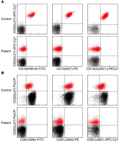
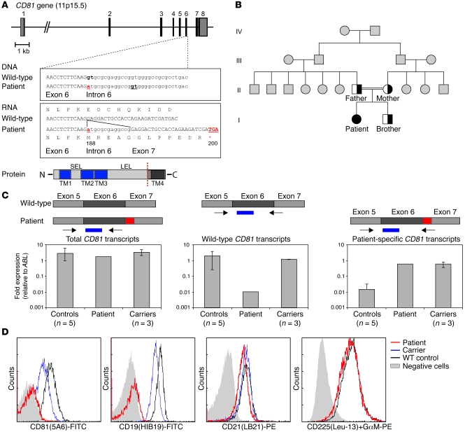
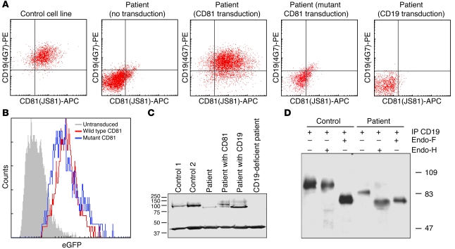
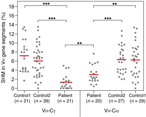
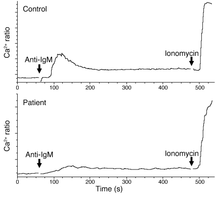
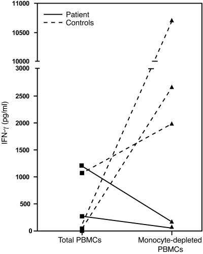
Antibody deficiencies constitute the largest group of symptomatic primary immunodeficiency diseases. In several patients, mutations in CD19 have been found to underlie disease, demonstrating the critical role for the protein encoded by this gene in antibody responses; CD19 functions in a complex with CD21, CD81, and CD225 to signal with the B cell receptor upon antigen recognition. We report here a patient with severe nephropathy and profound hypogammaglobulinemia. The immunodeficiency was characterized by decreased memory B cell numbers, impaired specific antibody responses, and an absence of CD19 expression on B cells. The patient had normal CD19 alleles but carried a homozygous CD81 mutation resulting in a complete lack of CD81 expression on blood leukocytes. Retroviral transduction and glycosylation experiments on EBV-transformed B cells from the patient revealed that CD19 membrane expression critically depended on CD81. Similar to CD19-deficient patients, CD81-deficient patients had B cells that showed impaired activation upon stimulation via the B cell antigen receptor but no overt T cell subset or function defects. In this study, we present what we believe to be the first antibody deficiency syndrome caused by a mutation in the CD81 gene and consequent disruption of the CD19 complex on B cells. These findings may contribute to unraveling the genetic basis of antibody deficiency syndromes and the nonredundant functions of CD81 in humans.


Figures

Figure 1
Figure 1. The patient’s B cells lack CD19 and CD81 membrane expression.
(A). Flow cytometric analysis of blood lymphocytes shows the complete absence of CD19 expression on CD20+ B cells from the patient. (B). The patient’s lymphocytes also completely lack CD81 membrane expression. In all panels, B cells are shown in red and other lymphocytes (mainly T cells and some NK cells) in black. The expression of both CD19 and CD81 was determined with 3 different monoclonal antibodies.
Figure 2
Figure 2. Homozygous CD81 splice site mutation results in alternative splicing and disruption of the CD19 complex on B cells.
(A). Schematic representation of the CD81 gene, consisting of 8 exons. The patient was homozygous for a splice site mutation downstream of exon 6 (exon6+1 G>A) resulting in the use of a cryptic splice site 13 nucleotides downstream of exon 6. The 13-nucleotide insertion results in a frameshift and a premature stop codon upstream of the fourth transmembrane domain of the CD81 protein. TM, transmembrane domain; SEL, short extracellular loop; LEL, large extracellular loop. (B). Pedigree of the family of the CD81-deficient patient. Half-filled symbols denote known carriers of the mutation; the filled symbol represents the patient, who is homozygous for the mutation; gray symbols denote family members who were not tested for carriership of the mutation; squares denote male family members; circles denote female family members. The parents of the patient were consanguineous (double line). (C). Quantitative analysis of CD81 transcripts in blood mononuclear cells. Three PCR assays were developed to quantify total, wild-type, and alternatively spliced CD81 transcripts. Arrows indicate primers; blue bars denote TaqMan probes. Data represent mean ± SD. (D). Expression levels of CD19 complex members on B cells of the patient and a carrier of the CD81 mutation. CD19 and CD81 expression were absent on patient’s B cells and reduced in carriers of the mutation. CD21 and CD225 were normally expressed. Isotype controls are shown for CD81 and CD225 stains and CD20-negative lymphocytes for CD19 and CD21 stains.
Figure 3
Figure 3. Loss of both CD19 and CD81 membrane expression on the patient’s B cells is due to the CD81 gene defect.
(A) The expression levels of CD19 and CD81 on EBV-transformed cell lines of the patient, a wild-type control, and patient cells after transduction with wild-type CD81, mutant CD81 (p.Glu188MetfsX13), or wild-type CD19. (B) eGFP expression levels in the patient’s EBV cells indicate similar expression of the wild-type and mutant CD81 constructs. (C) CD19 Western blot of EBV cell lines shows drastically reduced CD19 expression in the patient’s B cells. The molecular mass (Mr) of CD19 is lower than in controls and could only be restored upon transduction of CD81 and not CD19. (D) Immunoprecipitation followed by digestion with endo-H or endo-F demonstrated that the CD19 molecules in control and patient cells are sensitive to endo-F, and the deglycosylated CD19 molecules migrate similarly. However, only the patient’s CD19 is sensitive to endo-H digestion, indicating an ER/pre-Golgi location.
Figure 4
Figure 4. Reduced SHM in Vh gene segments of Cα and Cγ transcripts.
Sequence analysis revealed significantly reduced SHMs in Vh-Cα and Vh-Cγ transcripts of the patient as compared with age-matched controls. The frequency of mutations was significantly lower in Vh-Cγ transcripts of the patient as compared with Vh-Cα transcripts. Individual data points are shown, with red lines denoting mean values. **P < 0.01, ***P < 0.001.
Figure 5
Figure 5. Patient’s B cells show an intrinsic defect in signaling upon BCR stimulation.
The stimulation of blood mononuclear cells with anti-IgM F(ab′)2 fragments did not result in an initial calcium influx in CD20+ B cells from the patient, in contrast to control cells. The subsequent sustained influx of extracellular calcium was normal. No defect in calcium flux was seen in carriers of the CD81 mutation (data not shown). Ionomycin was added as a control for intracellular loading of Indo-1.
Figure 6
Figure 6. Tetanus toxoid–induced IFN-γ production by total PBMCs compared with monocyte-depleted PBMCs.
IFN-γ concentrations were measured by ELISA in the supernatants after 7-day cultures of blood mononuclear cells or monocyte-depleted cell suspensions in the presence of 5 μg/ml tetanus toxoid. In contrast to 3 controls, the patient’s responses were decreased when monocytes were depleted before culture. Dotted lines denote healthy controls, and solid lines represent the patient before (lower line) and 1 month after a tetanus toxoid booster (upper line).

Similar articles

Cited by

References

    1. Pan-Hammarstrom Q, Hammarstrom L. Antibody deficiency diseases. Eur J Immunol. 2008;38(2):327–333. - PubMed
    1. Conley ME, et al. Primary B cell immunodeficiencies: comparisons and contrasts. Annu Rev Immunol. 2009;27:199–227. doi: 10.1146/annurev.immunol.021908.132649. - DOI - PubMed
    1. Geha RS, et al. Primary immunodeficiency diseases: an update from the International Union of Immunological Societies Primary Immunodeficiency Diseases Classification Committee. J Allergy Clin Immunol. 2007;120(4):776–794. doi: 10.1016/j.jaci.2007.08.053. - DOI - PMC - PubMed
    1. Grimbacher B, et al. Homozygous loss of ICOS is associated with adult-onset common variable immunodeficiency. Nat Immunol. 2003;4(3):261–268. doi: 10.1038/ni902. - DOI - PubMed
    1. van Zelm MC, et al. An antibody-deficiency syndrome due to mutations in the CD19 gene. N Engl J Med. 2006;354(18):1901–1912. doi: 10.1056/NEJMoa051568. - DOI - PubMed

Publication types