. 2010 Apr;120(4):1151-64.
doi: 10.1172/JCI37223. Epub 2010 Mar 8.

Neutrophils responsive to endogenous IFN-beta regulate tumor angiogenesis and growth in a mouse tumor model

Affiliations

Neutrophils responsive to endogenous IFN-beta regulate tumor angiogenesis and growth in a mouse tumor model

Jadwiga Jablonska et al. J Clin Invest. 2010 Apr.

Erratum in

  • J Clin Invest. 2010 Nov 1;120(11):4163

Abstract

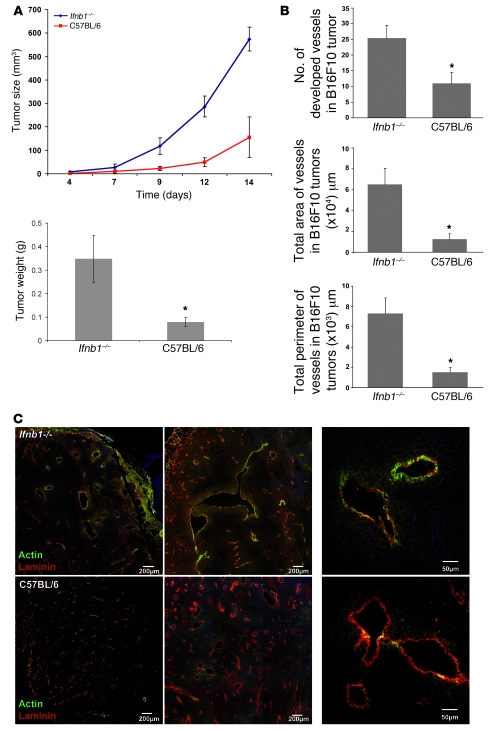
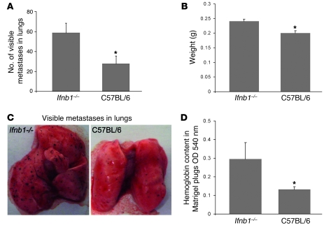
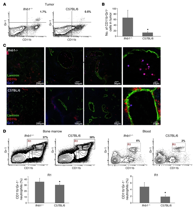
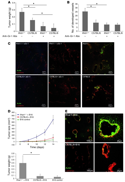
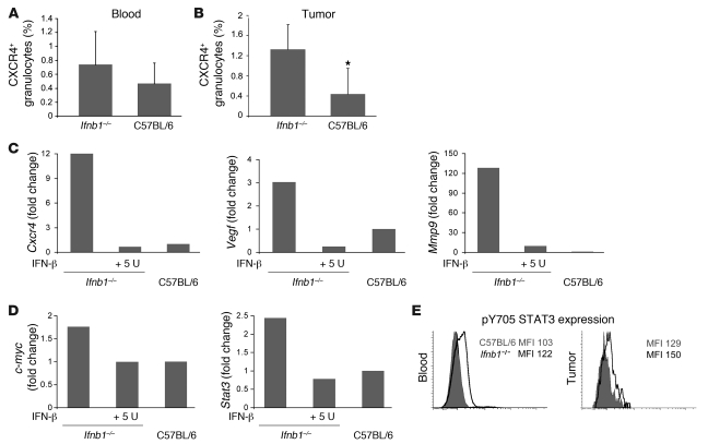
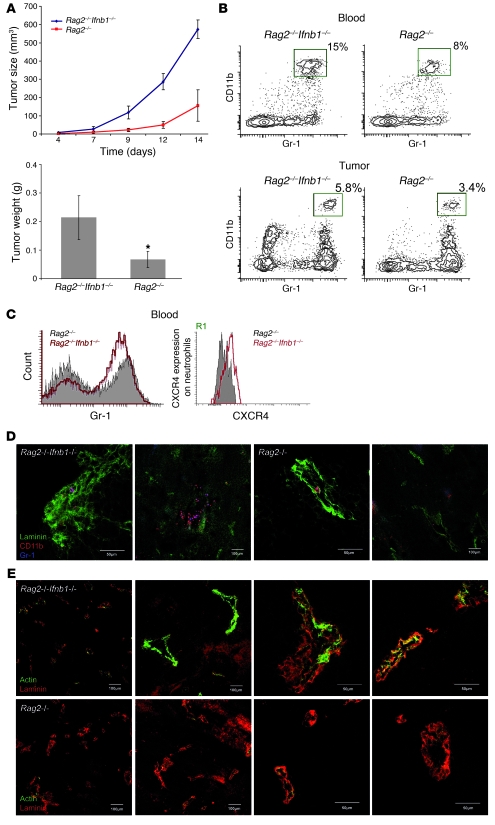
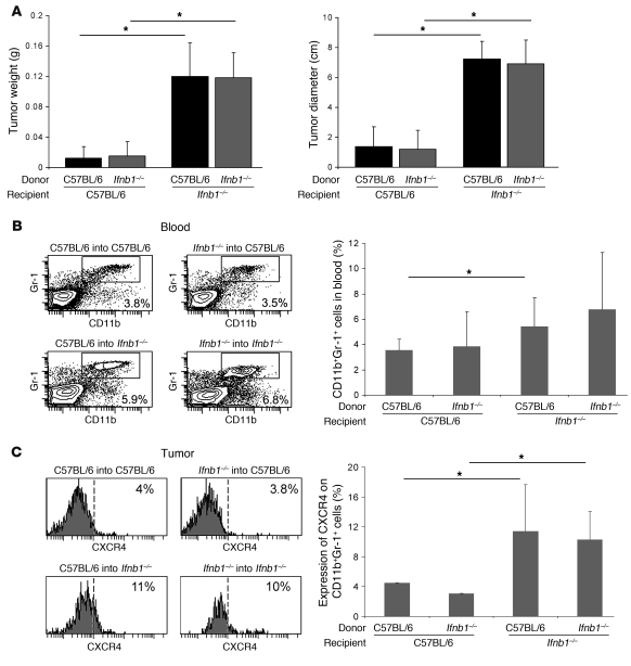
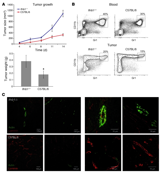
Angiogenesis is a hallmark of malignant neoplasias, as the formation of new blood vessels is required for tumors to acquire oxygen and nutrients essential for their continued growth and metastasis. However, the signaling pathways leading to tumor vascularization are not fully understood. Here, using a transplantable mouse tumor model, we have demonstrated that endogenous IFN-beta inhibits tumor angiogenesis through repression of genes encoding proangiogenic and homing factors in tumor-infiltrating neutrophils. We determined that IFN-beta-deficient mice injected with B16F10 melanoma or MCA205 fibrosarcoma cells developed faster-growing tumors with better-developed blood vessels than did syngeneic control mice. These tumors displayed enhanced infiltration by CD11b+Gr1+ neutrophils expressing elevated levels of the genes encoding the proangiogenic factors VEGF and MMP9 and the homing receptor CXCR4. They also expressed higher levels of the transcription factors c-myc and STAT3, known regulators of VEGF, MMP9, and CXCR4. In vitro, treatment of these tumor-infiltrating neutrophils with low levels of IFN-beta restored expression of proangiogenic factors to control levels. Moreover, depletion of these neutrophils inhibited tumor growth in both control and IFN-beta-deficient mice. We therefore suggest that constitutively produced endogenous IFN-beta is an important mediator of innate tumor surveillance. Further, we believe our data help to explain the therapeutic effect of IFN treatment during the early stages of cancer development.


Figures

Figure 1
Figure 1. Enhanced tumor growth and angiogenesis in Ifnb1–/– mice.
(A) Growth and size of tumors are significantly higher in Ifnb1–/– mice. B16F10 melanoma cells were injected s.c. into the abdomen of C57BL/6 or Ifnb1–/– mice, and tumor growth was monitored. At day 14, mice were sacrificed and tumor weight and diameter were measured. Experiments were done with at least 5 animals per group and repeated at least 3 times with similar results. Data represent mean ± SEM. *P ≤ 0.01. (B) The number, the area, and the perimeter of vessels in tumors isolated from Ifnb1–/– mice are significantly higher than in C57BL/6 mice. For histological analysis, material was collected as described in A. 10 μm cryosections were prepared and stained for laminin and actin (SMA). Fully developed vessels (laminin+actin+) were counted and the size of vessels calculated using ImageJ software. Histograms represent data from at least 3 independent experiments with at least 3 mice per group. More than 20 fields of view were analyzed in each experiment. Data represent mean ± SEM. *P ≤ 0.01. (C) B16F10 tumors grown in Ifnb1–/– mice exhibit a higher content of fully developed vessels (laminin+actin+). Histological analysis was done with material collected as described in A, with 10 μm cryosections prepared and stained for laminin (red) and actin (green). Scale bars: 200 μm and 50 μm, respectively. Photographs represent data from at least 3 independent experiments, with at least 3 mice per group.
Figure 2
Figure 2. Increased numbers of lung metastases and enhanced Matrigel angiogenesis in Ifnb1–/– mice.
(AC) Increased formation of metastases in lungs of Ifnb1–/– mice. Ifnb1–/– and control mice were injected i.v. with 4 × 10⁵ B16F10 melanoma cells. Mice were sacrificed after 14 days; lungs were removed and analyzed by counting visible B16F10 colonies and determining the weight of the lungs. (A) Number of visible colonies in lungs of Ifnb–/– and C57BL/6 mice. (B) Weight of lungs isolated from Ifnb1–/– and C57BL/6 mice. (C) Macroscopic comparison of lungs with B16F10 colonies from Ifnb1–/– and C57BL/6 mice. (D) In vivo angiogenesis assay shows higher vessel development in plugs isolated from Ifnb1–/– mice. BD Matrigel was injected s.c. into the abdomen of mice. At day 10, plugs were removed and analyzed for hemoglobin content using Drabkin reagent. Experiments were done twice, with at least 5 animals per group. Data represent mean ± SEM. *P ≤ 0.01.
Figure 3
Figure 3. Enhanced numbers of CD11b+Gr-1+ neutrophils in tumors of Ifnb1–/– mice.
(A) Percentage of CD11b+Gr-1+ cells (characterized in more detail in Supplemental Figure 1) in tumor is altered in Ifnb1–/– mice compared with C57BL/6 mice. Flow cytometry of myeloid cells from tumors of Ifnb1–/– and C57BL/6 mice. Tumors were removed at day 14; single-cell solutions were prepared, stained, and analyzed using the BD LSR II System. Data were analyzed with FACSDiva software. (B and C) Number of myeloid cells in tumors isolated from Ifnb1–/– mice is higher than in C57BL/6 mice. Tumors were removed, and 10 μm cryosections were prepared and stained for Gr-1 (blue), CD11b (red), and laminin (green). Number of neutrophils was calculated per 20 fields of view. Experiments were repeated at least twice, with at least 5 animals per group. Scale bars: 200 μm, 100 μm, 50 μm, and 20 μm respectively. (D) Percentage and number of CD11b+Gr-1+ neutrophils in bone marrow and blood are altered in Ifnb1–/– mice compared with C57BL/6 mice. Experiments were done twice with at least 5 animals per group. Data represent mean ± SEM. *P ≤ 0.01.
Figure 4
Figure 4. Essential role of IFN-β–responsive CD11b+Gr1+ neutrophils in B16F10 tumor growth and angiogenesis.
CD11b+Gr1+ cells were depleted by treatment with anti-Gr1 Ab, B16F10 cells injected s.c., and after 14 days, mice were sacrificed and tumors were removed, their weight determined, and cryosections stained for confocal microscopy. (A) Reduced tumor growth in mice depleted of CD11b+Gr1+ cells compared with untreated animals. (B and C) Number of developed vessels is reduced after depletion of Gr1+ cells. (D) Enhanced tumor growth depends strictly on type I IFN-reactive CD11b+Gr1+ neutrophils. Mice injected s.c. with B16F10 cells mixed with neutrophils obtained from tumor-bearing Ifnar1–/– mice (Ifnar–/–+B16) show increased tumor development compared with mice injected with neutrophils obtained from tumor-bearing WT mice (C57BL/6+B16) or B16 alone (B16 control). Experiments were carried out twice with at least 5 animals per group. Data represent mean ± SEM. *P ≤ 0.01. Histology shows representative pictures. At least 20 fields of view were analyzed. (E) B16F10 tumors coinjected with neutrophils obtained from tumor-bearing Ifnar1–/– mice (Ifnar–/–+B16) show a higher content of fully developed vessels (actin+laminin+) compared with B16F10 injected together with neutrophils obtained from tumor-bearing control mice (C57BL/6+B16).
Figure 5
Figure 5. Altered characteristics of CD11b+Gr1+ neutrophils isolated from Ifnb1–/– mice.
(A) Percentage of CXCR4+ neutrophils in blood of tumor-bearing Ifnb1–/– and C57BL/6 mice. (B) Percentage of CXCR4+ neutrophils isolated from tumors of Ifnb1–/– and C57BL/6 mice. Tumors were removed; single-cell solutions were prepared, stained, and analyzed using the BD LSR II System. Data were analyzed with FACSDiva software. Experiments were done twice with at least 5 animals per group. Data represent mean ± SEM. *P ≤ 0.01. (C) IFN-β treatment downregulates Vegf, Mmp9, and Cxcr4 gene expression. Tumors were removed; single-cell solutions were prepared and stained, and CD11b+Gr1+ neutrophils were isolated. Monolayers of such cells were incubated with 5 U rmIFN-β; after 4 hours, RNA was isolated, cDNA prepared, and gene expression measured using real-time RT-PCR. Cells were derived from 5 pooled tumors. All experiments were repeated at least 2 times. (D) High expression of Cxcr4 correlates with higher expression of c-myc and Stat3, and rmIFN-β downregulates both c-myc and Stat3. CD11b+Gr-1+ neutrophils were sorted from tumors and placed in culture with 5 U rmIFN-β. After 4 hours, cDNA was prepared as described in C. Cells were sorted from 5 pooled tumors. All experiments were repeated at least 2 times. (E) Expression of intracellular pSTAT3 in blood and tumor-infiltrating neutrophils was compared between control and Ifnb1–/– mice. Cell suspensions from blood and tumors were prepared, stained, and analyzed using the BD LSR II System. Data were analyzed with FACSDiva software. Experiments were done twice with at least 5 animals per group.
Figure 6
Figure 6. Restriction of tumor growth does not depend on T and B lymphocytes.
(A) Tumor growth in Rag2–/– mice is reduced compared with that in Rag2–/–Ifnb1–/– double-deficient mice. Tumor growth was monitored 14 days, and after this time, tumors removed. (B and C) Percentage and number of CD11b+Gr1+ neutrophils in blood and infiltrating tumors are higher in Rag2–/–Ifnb–/– mice. CXCR4 expression on blood neutrophils (R1 gate) is significantly increased in Rag2–/–Ifnb1–/– mice, which has an impact on their migration into tumor. (D) Immunohistochemistry of tumors isolated from Rag2–/–Ifnb1–/– and Rag2–/– mice shows higher number of infiltrating neutrophils in Rag2–/–Ifnb–/– mice compared with Rag2–/– animals. (E) Immunohistochemistry of tumors isolated from Rag2–/–Ifnb1–/– and Rag2–/– mice shows advanced angiogenic processes in Rag2–/–Ifnb1–/– mice compared with Rag2–/– animals. All experiments were carried out as above and done 3 times with at least 5 animals per group. Data represent mean ± SEM *P ≤ 0.01
Figure 7
Figure 7. IFN-β–producing cells are of radio-resistant, nonhematopoietic origin.
Recipient Ifnb1–/– and C57BL/6 mice were lethally irradiated and reconstituted with bone marrow from donor Ifnb1–/– or C57BL/6 mice. After 6 weeks, chimeras were injected s.c. with B16F10 melanoma cells. Tumors were removed at day 14 and analyzed. (A) Weight and diameter of isolated tumors. (B) Percentage of CD11b+Gr1+ neutrophils in blood. (C) Percentage of CXCR4+ cells in tumor-infiltrating CD11b+Gr1+ myeloid cell population. Experiments were done twice with at least 5 animals per group. Data represent mean ± SEM. *P ≤ 0.05.
Figure 8
Figure 8. Enhanced MCA205 fibrosarcoma growth and angiogenesis in Ifnb1–/– mice.
(A) Growth and size of tumors is significantly higher in Ifnb1–/– mice. MCA205 fibrosarcoma cells were injected s.c. into the abdomen of C57BL/6 or Ifnb1–/– mice, and tumor growth was monitored. At day 14, mice were sacrificed and tumor weight and diameter were measured. Experiments were done with at least 5 animals per group and repeated at least 3 times with similar results. Data represent mean ± SEM. *P ≤ 0.01. (B) Percentage of CD11b+Gr1+ neutrophils in blood and in Ifnb1–/– tumor-bearing mice is higher compared with the control. Tumors were removed at day 14; single-cell solutions prepared, stained, and analyzed using the BD LSR II System. Data were analyzed with FACSDiva software. (C) B16F10 tumors grown in Ifnb1–/– mice exhibit a higher content of fully developed vessels (laminin+actin+). Histological analysis was done with material collected as described in A, with 10-μm cryosections prepared and stained for laminin (red) and actin (green). Scale bars: 100 μm and 50 μm, respectively. Photographs represent data from at least 3 independent experiments, with at least 3 mice per group.

Similar articles

Cited by

References

    1. Folkman J. What is the evidence that tumors are angiogenesis dependent? J Natl Cancer Inst. 1990;82(1):4–6. - PubMed
    1. Plate KH, Breier G, Risau W. Molecular mechanisms of developmental and tumor angiogenesis. Brain Pathol. 1994;4(3):207–218. doi: 10.1111/j.1750-3639.1994.tb00835.x. - DOI - PubMed
    1. Auguste P, Lemiere S, Larrieu-Lahargue F, Bikfalvi A. Molecular mechanisms of tumor vascularization. Crit Rev Oncol Hematol. 2005;54(1):53–61. doi: 10.1016/j.critrevonc.2004.11.006. - DOI - PubMed
    1. Folkman J. Angiogenesis in cancer, vascular, rheumatoid and other disease. Nat Med. 1995;1(1):27–31. doi: 10.1038/nm0195-27. - DOI - PubMed
    1. Hanahan D, Weinberg RA. The hallmarks of cancer. Cell. 2000;100(1):57–70. doi: 10.1016/S0092-8674(00)81683-9. - DOI - PubMed

Publication types