

Review
doi: 10.4161/cc.9.6.10982.
Epub 2010 Mar 15.
Chronicles of a death foretold: dual sequential cell death checkpoints in TNF signaling
Affiliations
- PMID: 20237426
- PMCID: PMC4114106
- DOI: 10.4161/cc.9.6.10982
Item in Clipboard
Review
Chronicles of a death foretold: dual sequential cell death checkpoints in TNF signaling
Cell Cycle.
.
Abstract
The kinase RIP1 wears a coat of many colors during TNF receptor signaling and can regulate both activation of pro-survival NFkB and programmed cell death pathways. In this review, we outline how coating RIP1 with K63-linked ubiquitin chains forms a protective layer that prevents RIP1 from binding apoptotic regulators and serves as an early guard against cell death. Further on, binding of NFkB signaling components to the ubiquitin coat of RIP1 activates long-term pro-survival signaling and forms a more impenetrable suit of armor against cell death. If RIP1 is not decorated with ubiquitin chains it becomes an unstoppable harbinger of bad news: programmed cell death.
Figures
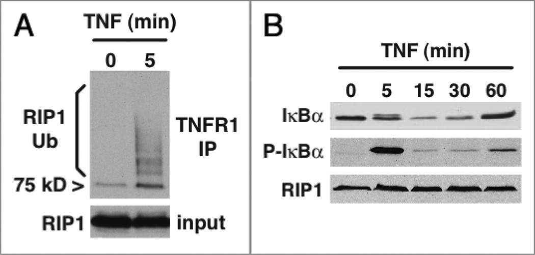
TNF induces rapid ubiquitination of RIP1. (A) Jurkat T cells were stimulated with 100 ng/ml human TNF for 5 minutes and TNFR1 was immunoprecipitated from the cytoplasmic protein fraction. The TNFR1 complex and a sample of the cytoplasmic lysate were probed by western blot for RIP1. The TNFR1 complex recruits the 75 kD RIP1 protein but the majority of the receptor-associated RIP1 undergoes modification consistent with ubiquitination (RIP1 Ub). (B) Jurkat T cells were stimulated with 100 ng/ml human TNF and cytoplasmic protein samples were probed by western blot for phosphorylated IκBα, total I㮫α and RIP1 as a loading control. Evidence of IKK activity is seen 5 minutes post-stimulation by the appearance of phosphorylated IκBα, co-incident with the polyubiquitination of receptor-associated RIP1 shown in (A). IκBα is then degraded but re-expression of this immediate early NFκB-inducible gene is not observable at the protein level until approximately one hour post stimulation. Therefore, the potentially pro-death RIP1 protein is bound to TNFR1 during the first hour of TNF stimulation before the appearance of NFκB-dependent gene products.
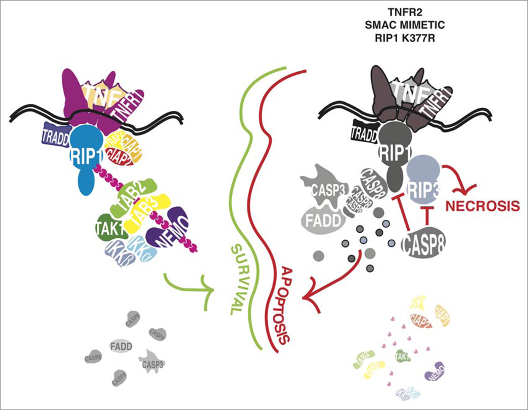
Ubiquitination of RIP1 is an early pro-survival checkpoint in TNF signaling. TNFR1 ligation triggers formation of a complex of TRADD, RIP1, TRAF2, cIAP1 and cIAP2 and the latter three enzymes catalyse the formation of K63-linked polyubiquitin chains on lysine 377 of RIP1. The K63-linked polyubiquitin chains form a docking site for proteins that contain a ubiquitin recognition motif such as TAB2, TAB3 and NEMO. These adaptor proteins recruit and activate the TAK1 and IKK complexes, which lead to phosphorylation and degradation of IκB. Formation of this complex on the ubiquitinated RIP1 prevents RIP1 from interacting with Caspase 8 and activating the apoptotic machinery. If the lysine 377 ubiquitin acceptor site of RIP1 is mutated or the E3 ligase enzymes cIAP1 and cIAP2 are degraded by treatment with SMAC mimetic or through TNFR2 ligation, non-ubiquitinated RIP1 rapidly forms a complex with Caspase 8 and turns on the apoptotic machinery. RIP1 and RIP3 are themselves substrates for Caspase 8, which may prevent them from triggering the alternate death pathway of programmed necrosis. If the activity of Caspase 8 is inhibited, RIP1 forms a complex with RIP3 and triggers cell death by necrosis, which is dependent on the kinase activity of RIP1 and RIP3. While binding of NEMO to ubiquitinated RIP1 is an important factor in this early pro-survival event, it remains unclear what contribution the binding of TAB2 and TAB3 or other proteins with a ubiquitin recognition domain might make in this pro-survival step. Similarly, it remains to be seen whether the TAK1 and IKK kinase complexes participate in a transcription-independent manner in this early pro-survival checkpoint, prior to long-term anti-death gene expression programs coming into effect.
Similar articles
-
Cellular IAP proteins and LUBAC differentially regulate necrosome-associated RIP1 ubiquitination.Cell Death Dis. 2015 Jun 25;6(6):e1800. doi: 10.1038/cddis.2015.158. Cell Death Dis. 2015. PMID: 26111062 Free PMC article.
-
Ubiquitination of RIP1 regulates an NF-kappaB-independent cell-death switch in TNF signaling.Curr Biol. 2007 Mar 6;17(5):418-24. doi: 10.1016/j.cub.2007.01.027. Epub 2007 Feb 15. Curr Biol. 2007. PMID: 17306544 Free PMC article.
-
Diverse ubiquitin linkages regulate RIP kinases-mediated inflammatory and cell death signaling.Cell Death Differ. 2017 Jul;24(7):1160-1171. doi: 10.1038/cdd.2017.33. Epub 2017 May 5. Cell Death Differ. 2017. PMID: 28475174 Free PMC article. Review.
-
Coordinated ubiquitination and phosphorylation of RIP1 regulates necroptotic cell death.Cell Death Differ. 2017 Jan;24(1):26-37. doi: 10.1038/cdd.2016.78. Epub 2016 Aug 12. Cell Death Differ. 2017. PMID: 27518435 Free PMC article.
-
The role of the kinases RIP1 and RIP3 in TNF-induced necrosis.Sci Signal. 2010 Mar 30;3(115):re4. doi: 10.1126/scisignal.3115re4. Sci Signal. 2010. PMID: 20354226 Review.
Cited by
-
The role of TRADD in death receptor signaling.Cell Cycle. 2012 Mar 1;11(5):871-6. doi: 10.4161/cc.11.5.19300. Epub 2012 Mar 1. Cell Cycle. 2012. PMID: 22333735 Free PMC article. Review.
-
Ripoptocide - A Spark for Inflammation.Front Cell Dev Biol. 2019 Aug 13;7:163. doi: 10.3389/fcell.2019.00163. eCollection 2019. Front Cell Dev Biol. 2019. PMID: 31457011 Free PMC article. Review.
-
Immune dysregulation in SHARPIN-deficient mice is dependent on CYLD-mediated cell death.Proc Natl Acad Sci U S A. 2021 Dec 14;118(50):e2001602118. doi: 10.1073/pnas.2001602118. Proc Natl Acad Sci U S A. 2021. PMID: 34887354 Free PMC article.
-
Zfra is a small wizard in the mitochondrial apoptosis.Aging (Albany NY). 2010 Dec;2(12):1023-9. doi: 10.18632/aging.100263. Aging (Albany NY). 2010. PMID: 21212468 Free PMC article.
-
CYLD Proteolysis Protects Macrophages from TNF-Mediated Auto-necroptosis Induced by LPS and Licensed by Type I IFN.Cell Rep. 2016 Jun 14;15(11):2449-61. doi: 10.1016/j.celrep.2016.05.032. Epub 2016 Jun 2. Cell Rep. 2016. PMID: 27264187 Free PMC article.
References
-
- Natoli G, Costanzo A, Guido F, Moretti F, Levrero M. Apoptotic, non-apoptotic and anti-apoptotic pathways of tumor necrosis factor signalling. Biochem Pharmacol. 1998;56:915–920. - PubMed
-
- Wang CY, Mayo MW, Korneluk RG, Goeddel DV, Baldwin AS., Jr NFkappaB antiapoptosis: induction of TRAF1 and TRAF2 and c-IAP1 and c-IAP2 to suppress caspase-8 activation. Science. 1998;281:1680–1683. - PubMed
-
- Micheau O, Tschopp J. Induction of TNF receptor I-mediated apoptosis via two sequential signaling complexes. Cell. 2003;114:181–190. - PubMed
-
- Liu ZG, Hsu H, Goeddel DV, Karin M. Dissection of TNF receptor 1 effector functions: JNK activation is not linked to apoptosis while NFkappaB activation prevents cell death. Cell. 1996;87:565–576. - PubMed
Publication types
MeSH terms
Substances
Grants and funding
LinkOut - more resources
Full Text Sources
Research Materials
Miscellaneous