. 2010 May;3(3):260-9.
doi: 10.1038/mi.2010.10. Epub 2010 Mar 17.

Human primary gastric dendritic cells induce a Th1 response to H. pylori

Affiliations

Human primary gastric dendritic cells induce a Th1 response to H. pylori

D Bimczok et al. Mucosal Immunol. 2010 May.

Abstract

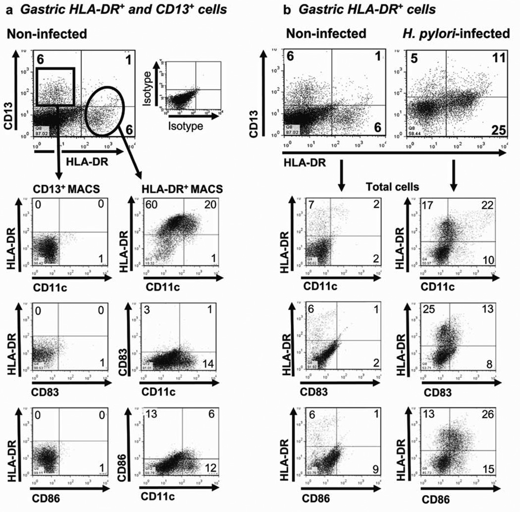
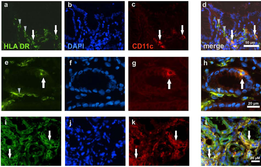
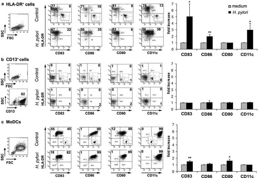
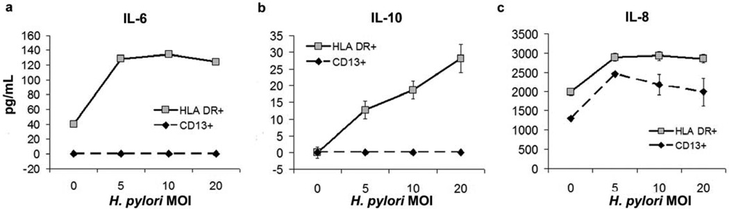
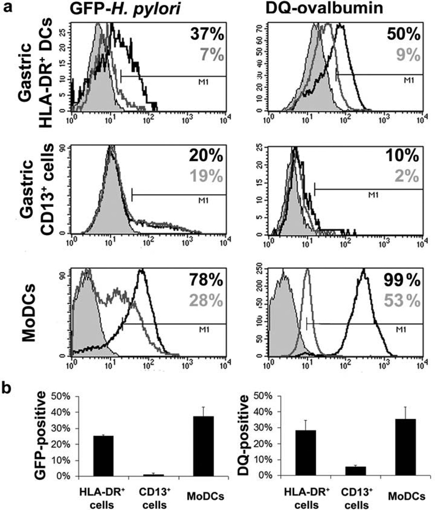
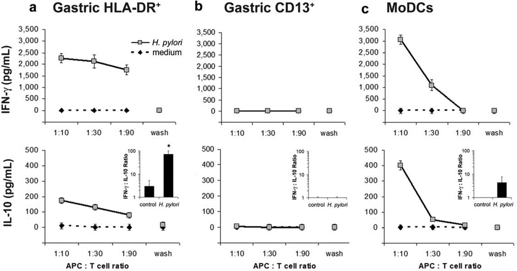
Adaptive CD4 T-cell responses are important in the pathogenesis of chronic Helicobacter pylori gastritis. However, the gastric antigen-presenting cells that induce these responses have not yet been identified. Here we show that dendritic cells (DCs) are present in the gastric mucosa of healthy subjects and are more prevalent and more activated in the gastric mucosa of H. pylori-infected subjects. H. pylori induced gastric DCs isolated from noninfected subjects to express increased levels of CD11c, CD86 and CD83, and to secrete proinflammatory cytokines, particularly interleukin (IL)-6 and IL-8. Importantly, gastric DCs pulsed with live H. pylori, but not control DCs, mediated T-cell secretion of interferon-gamma. The ability of H. pylori to induce gastric DC maturation and stimulate gastric DC activation of Th1 cells implicates gastric DCs as initiators of the immune response to H. pylori.


Conflict of interest statement

DISCLOSURE

The authors declared no conflicts of interest.

Figures

Figure 1
Figure 1
Phenotype of gastric mucosal cells from non-infected and H. pylori -infected subjects. (a) CD13+ and HLA-DR+ cells isolated from the gastric mucosa of a representative noninfected subject and enriched by magnetic antibody cell sorting (MACS) separation (n =10). (b) Phenotypic characterization of HLA-DR+ DCs among cells isolated from a representative, noninfected subject (n =4) and an H. pylori -infected subject (n =4). Numbers in the quadrants indicate percentage of cells in the respective quadrant.
Figure 2
Figure 2
Immunohistochemical identification of dendritic cells (DCs) in the gastric mucosa of noninfected and H. pylori -infected subjects. (a-d) Gastric mucosa from a noninfected subject contains HLA-DR+ cells in which a small proportion are HLADR+/ CD11c+. (e-h) Gastric mucosa from a noninfected subject shows gastric glands with an intraepithelial HLA-DR+/CD11c+ cell. (i-l) Gastric mucosa from an H. pylori infected subject contains many HLA-DR+ cells in which a large proportion are HLADR+/ CD11c+. Note the increased HLA-DR expression in gastric epithelial cells in the infected subject (i-l) compared with epithelial cells in the noninfected subject (a-h). (a,e,i) HLA-DR-FITC; (b,f,j) 46-diamidino-2-phenyl indole (DAPI) nuclear stain; (c,g,k) CD11c-TXRD (red); (d,h,l) merge. White arrows indicate HLA-DR+/CD11c+ DCs; gray arrowheads indicate HLA-DR+/CD11c cells, likely immature DCs. Results are representative of four noninfected and four H. pylori -infected subjects.
Figure 3
Figure 3
Maturation of gastric HLA-DR+ dendritic cells (DCs) and monocyte-derived DCs (MoDCs) in response to H. pylori. (a) HLA-DR+ DCs and (b) CD13+ cells isolated from the gastric mucosa of an H. pylori -negative donor, and (c) MoDCs generated from H. pylori -negative donor monocytes were exposed to live H. pylori (strain 60190, multiplicity of infection 20) or medium alone (control) for 15 h, followed by fluorescenceactivated cell sorting (FACS) analysis. Numbers in dot plots indicate percentage of cells in respective quadrants. Insets show isotype-matched controls. Bar graphs on the right show mean fold increase ± s.e.m. (n =3) in the expression of the indicated markers, calculated as f(x) = % stained cells (sample)/% stained cells (medium control); * P ≤0.05, ** P ≤0.01; unpaired Student’s t -test.
Figure 4
Figure 4
H. pylori -induced cytokine secretion by gastric dendritic cells (DCs). Culture supernatants of gastric HLA-DR+ DCs or CD13+ cells obtained from H. pylori -negative donors and stimulated with H. pylori in vitro were collected after 15 h, and secretion of (a) IL-6, (b) IL-10 and (c) IL-8 was determined by enzyme-linked immunosorbent assay. Data are from a representative experiment (n =3, using cells from separate subjects); values correspond to mean ± s.d.
Figure 5
Figure 5
Antigen uptake and processing by gastric HLA-DR+ dendritic cells (DCs), CD13+ cells and monocyte-derived DCs (MoDCs). Magnetic antibody cell sortingenriched gastric HLA-DR+ DCs or CD13+ cells or 5-day-old MoDCs from H. pylori negative donors were incubated for 30 min in the presence of GFP- H. pylori (multiplicity of infection 10; left panels) or DQ-ovalbumin (10 µg/mL; right panels) at 37 or 4°C. (a) Solid grey histograms correspond to cells incubated in medium alone, open grey histograms to cells along with GFP- H. pylori or DQ-ovalbumin at 4°C, and open black histograms to cells along with GFP- H. pylori or DQ-ovalbumin at 37°C. (b) Percentage of cells positive for GFP- H. pylori (left panel) and DQ-ovalbumin (right panel) expressed as mean ± s.e.m. of 2–3 independent experiments; values corrected for nonspecific binding at 4°C.
Figure 6
Figure 6
Gastric HLA-DR+ dendritic cells (DCs) drive a Th1-predominant response. (a) Gastric HLA-DR+ DCs, (b) gastric CD13+ cells or (c) monocyte-derived DCs (MoDCs) derived from cells from healthy subjects were pulsed with H. pylori, multiplicity of infection 20 for 2 h, washed twice, and co-cultured with autologous blood CD4+ T cells for 3 days. Supernatants from the second wash (“wash”) were used as controls to exclude direct effects of residual bacteria on the T cells. Concentrations of interfreron (IFN)-γ and interleukin (IL)-10 in culture supernatants were determined by enzymelinked immunosorbent assay. Data are representative of three (gastric HLA-DR+ DCs) or two (gastric CD13+ cells and MoDCs) experiments with cells from separate subjects; values correspond to mean ± s.e.m.. Insets in the lower panels show the ratio of IFN-γ to IL-10 for two or three experiments (mean ± s.e.m.) at an antigen-presenting cell (APC)/T-cell ratio of 1:10; * P <0.05, unpaired Student’s t -test.

Similar articles

Cited by

References

    1. Parkin DM. The global health burden of infection-associated cancers in the year 2002. Int. J. Cancer. 2006;118:3030–3044. - PubMed
    1. Peek RM, Jr, Blaser MJ. Helicobacter pylori and gastrointestinal tract adenocarcinomas. Nat. Rev. Cancer. 2002;2:28–37. - PubMed
    1. Bamford KB, et al. Lymphocytes in the human gastric mucosa during Helicobacter pylori have a T helper cell 1 phenotype. Gastroenterology. 1998;114:482–492. - PubMed
    1. Kraft M, et al. IFN-gamma synergizes with TNF-alpha but not with viable H. pylori in up-regulating CXC chemokine secretion in gastric epithelial cells. Clin. Exp. Immunol. 2001;126:474–481. - PMC - PubMed
    1. Kim JM, Kim JS, Jung HC, Song IS, Kim CY. Up-regulation of inducible nitric oxide synthase and nitric oxide in Helicobacter pylori -infected human gastric epithelial cells: possible role of interferon-gamma in polarized nitric oxide secretion. Helicobacter. 2002;7:116–128. - PubMed

Publication types