

Self-complementary AAV-mediated gene therapy restores cone function and prevents cone degeneration in two models of Rpe65 deficiency
- PMID: 20237510
- PMCID: PMC3014850
- DOI: 10.1038/gt.2010.29
Self-complementary AAV-mediated gene therapy restores cone function and prevents cone degeneration in two models of Rpe65 deficiency
Abstract
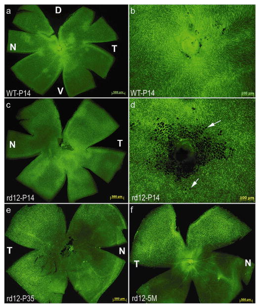
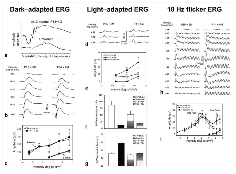
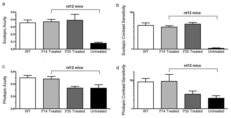
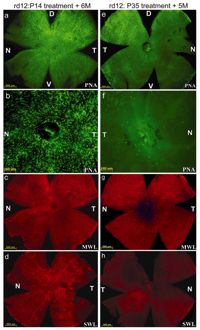
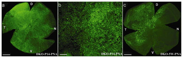
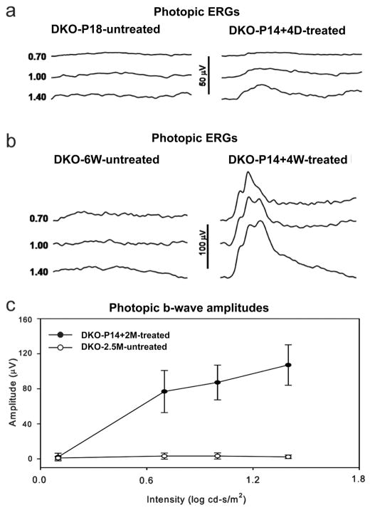
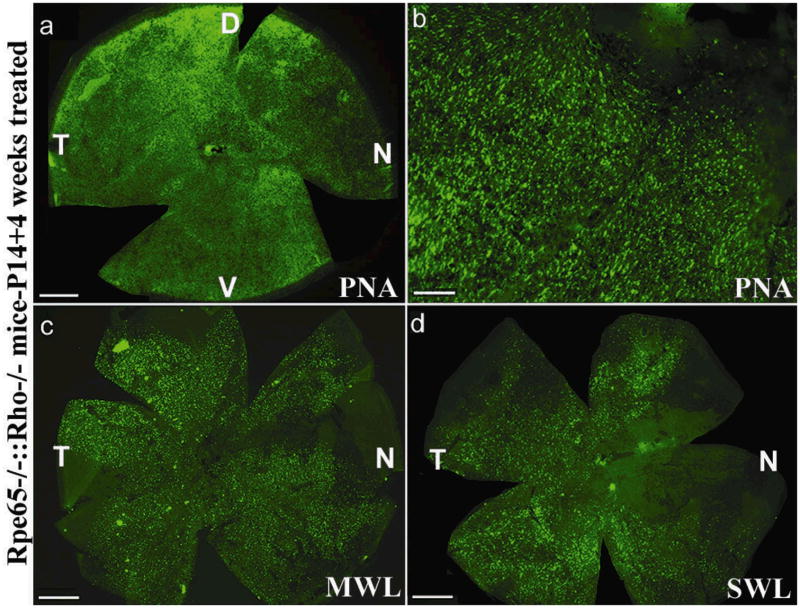
To test whether fast-acting, self-complimentary (sc), adeno-associated virus-mediated RPE65 expression prevents cone degeneration and/or restores cone function, we studied two mouse lines: the Rpe65-deficient rd12 mouse and the Rpe65-deficient, rhodopsin null ('that is, cone function-only') Rpe65(-/-)::Rho(-/-) mouse. scAAV5 expressing RPE65 was injected subretinally into one eye of rd12 and Rpe65(-/-)::Rho(-/-) mice at postnatal day 14 (P14). Contralateral rd12 eyes were injected later, at P35. Rd12 behavioral testing revealed that rod vision loss was prevented with either P14 or P35 treatment, whereas cone vision was only detected after P14 treatment. Consistent with this observation, P35 treatment only restored rod electroretinogram (ERG) signals, a result likely due to reduced cone densities at this time point. For Rpe65(-/-)::Rho(-/-) mice in which there is no confounding rod contribution to the ERG signal, cone cells and cone-mediated ERGs were also maintained with treatment at P14. This work establishes that a self-complimentary AAV5 vector can restore substantial visual function in two genetically distinct models of Rpe65 deficiency within 4 days of treatment. In addition, this therapy prevents cone degeneration but only if administered before extensive cone degeneration, thus supporting continuation of current Leber's congenital amaurosis-2 clinical trials with an added emphasis on cone subtype analysis and early intervention.
Figures
Similar articles
-
Gene therapy rescues cone structure and function in the 3-month-old rd12 mouse: a model for midcourse RPE65 leber congenital amaurosis.Invest Ophthalmol Vis Sci. 2011 Jan 5;52(1):7-15. doi: 10.1167/iovs.10-6138. Print 2011 Jan. Invest Ophthalmol Vis Sci. 2011. PMID: 21169527 Free PMC article.
-
Gene therapy following subretinal AAV5 vector delivery is not affected by a previous intravitreal AAV5 vector administration in the partner eye.Mol Vis. 2009;15:267-75. Epub 2009 Feb 6. Mol Vis. 2009. PMID: 19190735 Free PMC article.
-
Gene therapy restores vision-dependent behavior as well as retinal structure and function in a mouse model of RPE65 Leber congenital amaurosis.Mol Ther. 2006 Mar;13(3):565-72. doi: 10.1016/j.ymthe.2005.09.001. Epub 2005 Oct 11. Mol Ther. 2006. PMID: 16223604
-
RPE65: role in the visual cycle, human retinal disease, and gene therapy.Ophthalmic Genet. 2009 Jun;30(2):57-62. doi: 10.1080/13816810802626399. Ophthalmic Genet. 2009. PMID: 19373675 Free PMC article. Review.
-
Leber congenital amaurosis due to RPE65 mutations and its treatment with gene therapy.Prog Retin Eye Res. 2010 Sep;29(5):398-427. doi: 10.1016/j.preteyeres.2010.04.002. Epub 2010 Apr 24. Prog Retin Eye Res. 2010. PMID: 20399883 Free PMC article. Review.
Cited by
-
The Degeneration and Apoptosis Patterns of Cone Photoreceptors in rd11 Mice.J Ophthalmol. 2017;2017:9721362. doi: 10.1155/2017/9721362. Epub 2017 Jan 12. J Ophthalmol. 2017. PMID: 28168050 Free PMC article.
-
Gene delivery to the retina: from mouse to man.Methods Enzymol. 2012;507:255-74. doi: 10.1016/B978-0-12-386509-0.00013-2. Methods Enzymol. 2012. PMID: 22365778 Free PMC article.
-
The Lrat-/- Rat: CRISPR/Cas9 Construction and Phenotyping of a New Animal Model for Retinitis Pigmentosa.Int J Mol Sci. 2021 Jul 5;22(13):7234. doi: 10.3390/ijms22137234. Int J Mol Sci. 2021. PMID: 34281288 Free PMC article.
-
The frequency-response electroretinogram distinguishes cone and abnormal rod function in rd12 mice.PLoS One. 2015 Feb 23;10(2):e0117570. doi: 10.1371/journal.pone.0117570. eCollection 2015. PLoS One. 2015. PMID: 25706871 Free PMC article.
-
Advances in gene therapy technologies to treat retinitis pigmentosa.Clin Ophthalmol. 2014;8:127-36. doi: 10.2147/OPTH.S38041. Epub 2013 Dec 24. Clin Ophthalmol. 2014. PMID: 24391438 Free PMC article. Review.
References
-
- Marlhens F, Bareil C, Griffoin JM, Zrenner E, Amalric P, Eliaou C, et al. Mutations in RPE65 cause Leber’s congenital amaurosis. Nat Genet. 1997;17:139–41. - PubMed
-
- Rohrer B, Lohr HR, Humphries P, Redmond TM, Seeliger MW, Crouch RK. Cone opsin mislocalization in Rpe65−/− mice: a defect that can be corrected by 11-cis retinal. Invest Ophthalmol Vis Sci. 2005;46:3876–82. - PubMed
Publication types
MeSH terms
Substances
Grants and funding
- EY08571/EY/NEI NIH HHS/United States
- EY13729/EY/NEI NIH HHS/United States
- R01 EY019320/EY/NEI NIH HHS/United States
- P30 EY006360/EY/NEI NIH HHS/United States
- F32 EY017246/EY/NEI NIH HHS/United States
- EY07758/EY/NEI NIH HHS/United States
- P01 NS036302/NS/NINDS NIH HHS/United States
- EY017246/EY/NEI NIH HHS/United States
- NS36302/NS/NINDS NIH HHS/United States
- EY00067/EY/NEI NIH HHS/United States
- EY11123/EY/NEI NIH HHS/United States
- R21 EY023543/EY/NEI NIH HHS/United States
- P30 EY008571/EY/NEI NIH HHS/United States
- P30 EY021721/EY/NEI NIH HHS/United States
- U10 EY013729/EY/NEI NIH HHS/United States
- R01 EY007758/EY/NEI NIH HHS/United States
- R01 EY011123/EY/NEI NIH HHS/United States
- EY06360/EY/NEI NIH HHS/United States
- R01 EY019943/EY/NEI NIH HHS/United States
- EY014046/EY/NEI NIH HHS/United States
- EY018331/EY/NEI NIH HHS/United States
- R21 EY018331/EY/NEI NIH HHS/United States
LinkOut - more resources
Full Text Sources
Other Literature Sources
Medical
Research Materials
