. 2010 May;133(5):505-15.
doi: 10.1007/s00418-010-0683-0. Epub 2010 Mar 18.

Human LPLUNC1 is a secreted product of goblet cells and minor glands of the respiratory and upper aerodigestive tracts

Affiliations

Human LPLUNC1 is a secreted product of goblet cells and minor glands of the respiratory and upper aerodigestive tracts

Colin D Bingle et al. Histochem Cell Biol. 2010 May.

Abstract

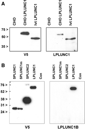
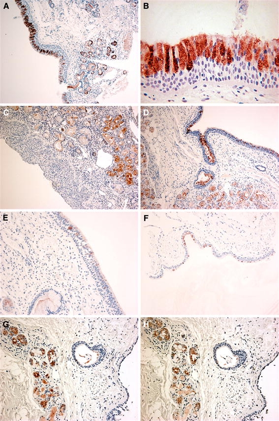
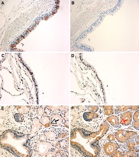
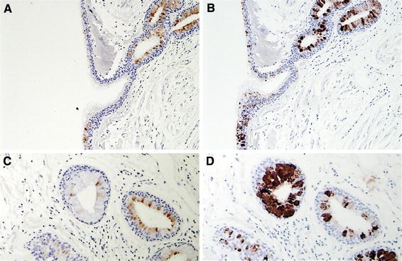
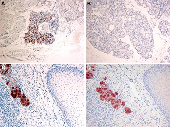
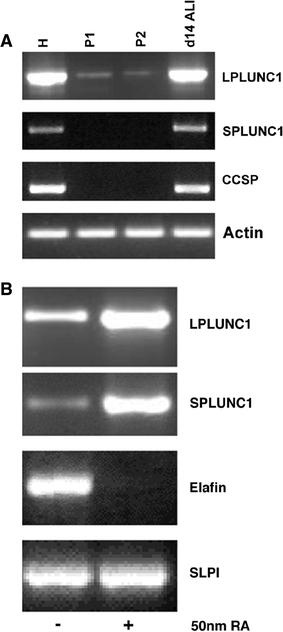
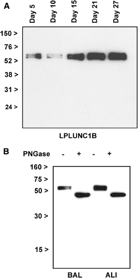
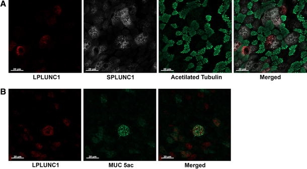
Long PLUNC1 (LPLUNC1, C20orf114) is a member of a family of poorly described proteins (PLUNCS) expressed in the upper respiratory tract and oral cavity, which may function in host defence. Although it is one of the most highly expressed genes in the upper airways and has been identified in sputum and nasal secretions by proteomic studies, localisation of LPLUNC1 protein has not yet been described. We developed affinity purified antibodies and localised the protein in tissues of the human respiratory tract, oro- and nasopharynx. We have complemented these studies with analysis of LPLUNC1 expression in primary human lung cell cultures and used Western blotting to study the protein in cell culture secretions and in BAL. LPLUNC1 is a product of a population of goblet cells in the airway epithelium and nasal passages and is also present in airway submucosal glands and minor glands of the oral and nasal cavities. The protein is not expressed in peripheral lung epithelial cells. LPLUNC1 is present in bronchoalveolar lavage fluid as two glycosylated isoforms and primary airway epithelial cells produce identical proteins as they undergo mucociliary differentiation. Our results suggest that LPLUNC1 is an abundant, secreted product of goblet cells and minor mucosal glands of the respiratory tract and oral cavity and suggest that the protein functions in the complex milieu that protects the mucosal surfaces in these locations.


Figures

Fig. 1
Fig. 1
Characterisation of the LPLUNC1 antibody by Western blotting. a V5-epitope tagged LPLUNC1 proteins were generated by coupled in vitro transcription and translation (Ivt-LPLUNC1) or from conditioned medium from stable CHO expressing cell lines (CHO LPLUNC1) or control medium (CHO) as outlined in “Materials and methods”. 2 μl of the IVT reaction or 5 μl of concentrated conditioned media was resolved on replicate 12% SDS-PAGE gels and Western blotted using the LPLUNC1B polyclonal antibody or an antibody to the V5 epitope as described. The black arrows indicate the positions of the molecular mass markers. b V5-epitope tagged PLUNC proteins were generated from conditioned medium from stable CHO expressing cell lines, expressing SPLUNC1 a cystine-mutant SPLUNC1 (SPLUNC1m), SPLUNC2 or LPLUNC1 or control medium (Con) as outlined in “Materials and methods”. 5 μl of concentrated conditioned media was resolved on replicate 12% SDS-PAGE gels and Western blotted using the LPLUNC1B polyclonal antibody or an antibody to the V5 epitope as described. The black arrows indicate the positions of the molecular mass markers
Fig. 2
Fig. 2
Distribution of LPLUNC1 in the respiratory tract. Immunohistochemistry for LPLUNC1 was performed with the LPLUNC1B antibody as described in “Materials and methods”. Sections show staining in samples of nasal antral mucosa (ac), trachea (d), bronchial airways (e) and bronchiolar epithelium (f). Serial sections of trachea were stained with both antibodies, LPLUNC1A (g) and LPLUNC1B (h). The original magnifications of the images were ≈×100 (a, c, d), ×200 (eh) and ×400 (b)
Fig. 3
Fig. 3
LPLUNC1 and SPLUNC1 do not co-localise in the respiratory tract. Immunohistochemistry was performed as described in “Materials and methods” for LPLUNC1 (ae) and SPLUNC1 (b, d, f). Sections show staining in serial samples of airways (ad) and in an airway submucosal gland (e, f). The black arrow in e shows a serous demilune that is positive for LPLUNC1 whereas the red arrow in f points to the same group of cells that is negative for SPLUNC1. The original magnifications of the images were ×200 (a, b) and ×400 (cf)
Fig. 4
Fig. 4
LPLUNC1 and MUC5A/C do not always co-localise in the respiratory tract. Immunohistochemistry was performed as described in “Materials and methods” for LPLUNC1 (a, c) and MUC5A/C (b, d). Sections show staining in serial samples of airways (a, d) and in an airway submucosal gland (c, d). The original magnifications of the images were ×200 (a, b) and ×400 (c, d)
Fig. 5
Fig. 5
Distribution of LPLUNC1 in minor glands of the oral cavity. Immunohistochemistry for LPLUNC1 and SPLUNC1 was performed as described in “Materials and methods”. Sections show staining in a seromucous gland of in the tongue (a) but no staining in a minor mucus gland in the proximal tongue (b). Serial sections of minor glands surrounding the palatine tonsils show that both LPLUNC1 (c) and SPLUNC1 (d) are positive in the same region. The original magnifications of the images were ×200 (a, b) and ×400 (c, d)
Fig. 6
Fig. 6
LPLUNC1 is expressed in primary tracheal cells and cells differentiated at the ALI. a Expression of LPLUNC1 was studied by PCR using freshly harvested human tracheal cells (H) and samples taken from the cultures at two stages (P1, P2) as the cells were established at an air–liquid interface (ALI). Expression of SPLUNC1, the non-ciliated tracheal cell marker CCSP and actin (as a PCR control) was also monitored in the same samples. b Expression of LPLUNC1 was studied by PCR in samples of primary tracheobronchial epithelial cells during retinoic acid depletion and the subsequent loss of the differentiated phenotype. ALI cells were established using standard growth conditions in medium containing 50 nM all trans retinoic acid (RA). After 14 days in culture RA was removed from the medium of one group of cells and culture was continued for an additional 18 days. Expression of LPLUNC1 and SPLUNC1 was investigated by RT-PCR with exon spanning primer pairs as described in “Materials and methods” with primers to SLPI and elafin serving as a positive controls. RT-negative PCR reactions were run for each experiment and representative RCR products were cloned and sequenced for confirmation. The data in both panels represents one of three sets of experiments performed with cells from different donors
Fig. 7
Fig. 7
LPLUNC1 is secreted into the apical fluid as a glycosylated protein identical to that found in BAL fluid. a Secretion of LPLUNC1 into apical secretions harvested from ALI cell cultures was shown by Western blotting using the LPLUNC1B antibody. Samples were harvested from the cells every 2–3 days. The positions of the molecular mass markers are indicated by the black arrows. The data is representative of two different experiments. b Deglycosylation studies were performed using BAL fluid and ALI secretions as described in “Materials and methods”. The resultant reaction products were subjected to Western blotting and detected with the LPLUNC1B antibody. The positions of the molecular mass markers are indicated by the black arrows
Fig. 8
Fig. 8
LPLUNC1 is localised in non-ciliated MUC5AC positive cells in TBE ALI cell cultures. Confocal IF microscopy was performed on differentiated ALI cultures as described in “Materials and methods” for LPLUNC1, SPLUNC1, tubulin and MUC5A/C as described. a LPLUNC1 and SPLUNC1 are localised in non-ciliated cells. Ciliated cells were identified with IgG anti-acetylated tubulin present in ciliary structures. The population of SPLUNC1-positive cells is more abundant than LPLUNC1 positive cells. b LPLUNC1 is present in some MUC5AC positive cells. All pictures were taken with a Zeiss LSM-510 confocal microscope, at ×40 with a ×2 digital zoom

Similar articles

Cited by

References

    1. Barnes FA, Bingle L, Bingle CD. Pulmonary genomics, proteomics and PLUNCs. Am J Respir Cell Mol Biol. 2008;38:377–379. doi: 10.1165/rcmb.2007-0388TR. - DOI - PubMed
    1. Beamer LJ, Carroll SF, Eisenberg D. Crystal structure of human BPI and two bound phospholipids at 2.4 angstrom resolution. Science. 1997;276:1861–1864. doi: 10.1126/science.276.5320.1861. - DOI - PubMed
    1. Bingle CD, Bingle L. Characterisation of the human plunc gene, a gene product with an upper airways and nasopharyngeal restricted expression pattern. Biochim Biophys Acta. 2000;1493:363–367. - PubMed
    1. Bingle CD, Craven CJ. PLUNC: a novel family of candidate host defence proteins expressed in the upper airways and nasopharynx. Hum Mol Genet. 2002;11:937–943. doi: 10.1093/hmg/11.8.937. - DOI - PubMed
    1. Bingle CD, Craven CJ. Meet the relatives: a family of BPI and LBP-related proteins. Trends Immunol. 2004;25:53–55. doi: 10.1016/j.it.2003.11.007. - DOI - PubMed

Publication types

MeSH terms