. 2010 Jul;67(13):2271-81.
doi: 10.1007/s00018-010-0331-9. Epub 2010 Mar 18.

Annexin A4 interacts with the NF-kappaB p50 subunit and modulates NF-kappaB transcriptional activity in a Ca2+-dependent manner

Affiliations

Annexin A4 interacts with the NF-kappaB p50 subunit and modulates NF-kappaB transcriptional activity in a Ca2+-dependent manner

Young-Joo Jeon et al. Cell Mol Life Sci. 2010 Jul.

Abstract

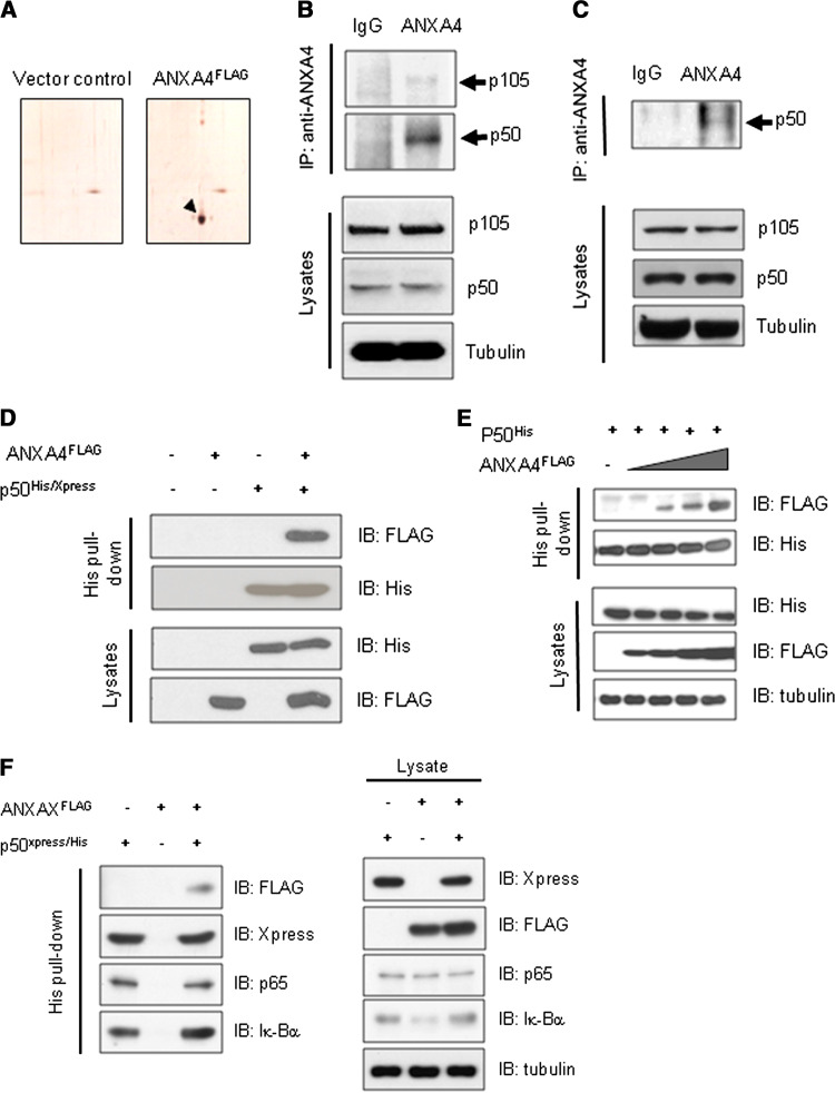
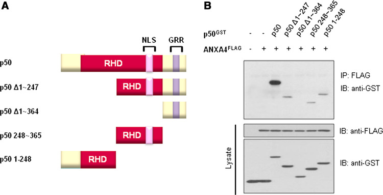
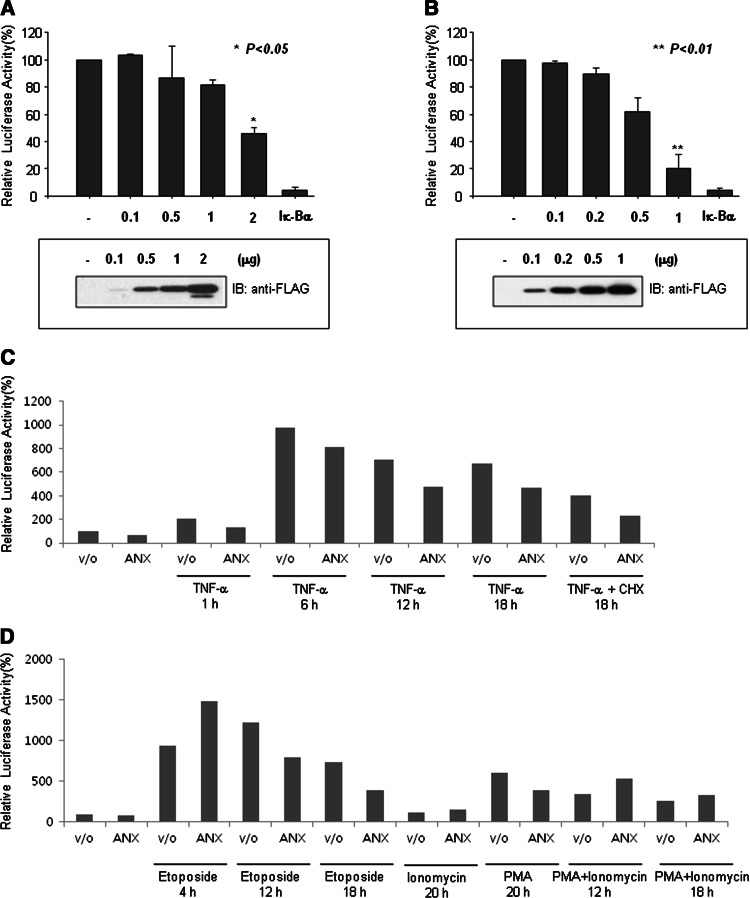
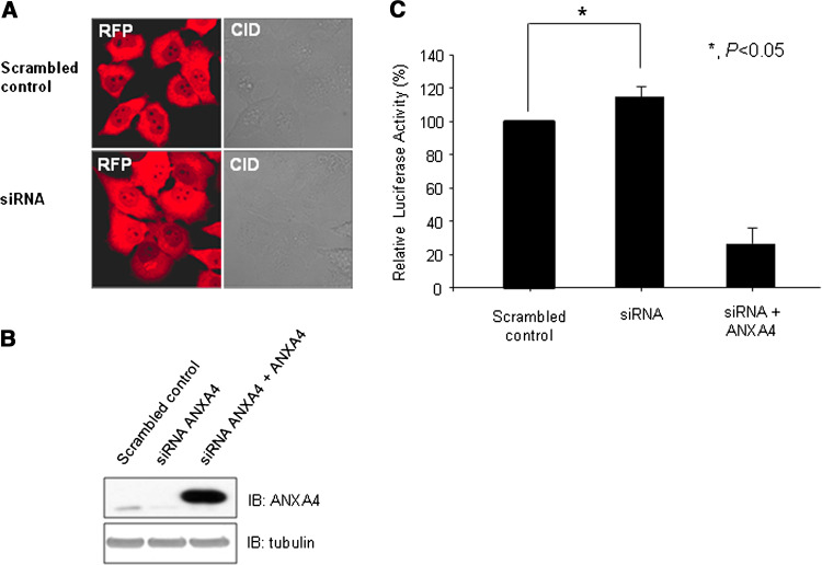
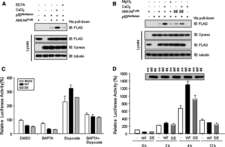
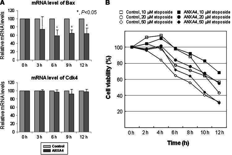
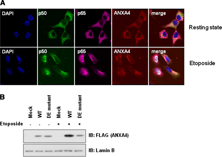
Previously, we identified annexin A4 (ANXA4) as a candidate substrate of caspase-3. Proteomic studies were performed to identify interacting proteins with a view to determining the roles of ANXA4. ANXA4 was found to interact with the p105. Subsequent studies revealed that ANXA4 interacts with NF-kappaB through the Rel homology domain of p50. Furthermore, the interaction is markedly increased by elevated Ca(2+) levels. NF-kappaB transcriptional activity assays demonstrated that ANXA4 suppresses NF-kappaB transcriptional activity in the resting state. Following treatment with TNF-alpha or PMA, ANXA4 also suppressed NF-kappaB transcriptional activity, which was upregulated significantly early after etoposide treatment. This difference may be due to the intracellular Ca(2+) level. Additionally, ANXA4 translocates to the nucleus together with p50, and imparts greater resistance to apoptotic stimulation by etoposide. Our results collectively indicate that ANXA4 differentially modulates the NF-kappaB signaling pathway, depending on its interactions with p50 and the intracellular Ca(2+) ion level.


Figures

Fig. 1
Fig. 1
ANXA4 interacts with p50 and p105, and has no effect on the composition of the NF-κB complex. a Enlarged image of a 2-DE gel showing the p105 spot in a control gel, compared to that in a gel presenting an ANXA4-transfectant. The detailed protocol is described in Materials and methods. b Cell lysates were prepared from Caki-1 cells that overexpress ANXA4 protein. Extracts were immunoprecipitated with anti-ANXA4 goat polyclonal IgG or anti-goat preimmune IgG, separated by SDS-PAGE, and analyzed by Western blotting with NF-κB p105/p50-specific antibody for simultaneous detection of both proteins. c Endogenous interactions between ANXA4 and p50 were assessed in HeLa cells. Extracts were immunoprecipitated with anti-ANXA4 goat polyclonal IgG or anti-goat preimmune IgG, separated by SDS-PAGE, and analyzed by Western blotting with p50-specific antibody. d HEK-293 cells were transfected with vectors containing FLAG-tagged ANXA4 and His/Xpress-tagged p50 genes, and extracted with Ni-NTA lysis buffer. Extracts were pulled down with Ni-NTA agarose beads and pellets analyzed by Western blotting. e Different amounts of FLAG-tagged ANXA4 DNA (0, 0.1, 0.5, 1, and 2 µg) were transfected into HEK-293 cells with His-tagged p50 DNA (1 µg). Transfected HEK-293 cells were extracted and pulled down using Ni-NTA agarose beads. Pellets were analyzed by Western blotting with anti-FLAG antibody. f FLAG-tagged ANXA4 DNA (1 µg) was transfected into HEK-293 cells with His/Xpress-tagged p50 DNA (1 µg). Transfected HEK-293 cells were extracted and pulled down using Ni-NTA agarose beads. Pellets were analyzed by Western blotting with anti-FLAG, anti-Xpress, p65, and Iκ-Bα antibodies
Fig. 2
Fig. 2
p50 interacts with ANXA4 through its Rel homology domain. a Structural map of p50 and several deletion mutant proteins used in this study are depicted; the Rel homology domain (RHD), nuclear localization sequence (NLS), and glycine-rich region (GRR) are designated. b To determine the region of p50 required for association with ANXA4, Bosc 23 cells were co-transfected with GST-tagged p50 truncation mutants and FLAG-tagged ANXA4. Transfected cells were subjected to GST pull-down assays, and analyzed by Western-blot analysis with the anti-FLAG antibody
Fig. 3
Fig. 3
NF-κB transcriptional activity assay after enforced expression of ANXA4. a The vector encoding C-terminal FLAG-tagged ANXA4 was transfected into HeLa cells at the indicated amounts and then luciferase assays were performed. Below the graph, the amounts of overexpressed ANXA4 proteins determined by Western blotting with an anti-FLAG antibody are shown. b The effect of C-terminal FLAG-tagged ANXA4 expression in HEK-293 cells on NF-κB transcription activity was investigated, as described in a. c The NF-κB transcriptional assay after TNF-α stimulation was performed in HeLa cells transfected with vector encoding C-terminal FLAG-tagged ANXA4. d The NF-κB transcriptional assay after etoposide or PMA stimulation was performed in HeLa cells transfected with a vector encoding C-terminal FLAG-tagged ANXA4. To ascertain whether Ca2+ modulates NF-κB transcriptional activity induced by ANXA4, cells were co-treated with ionomycin and PMA
Fig. 4
Fig. 4
NF-κB transcriptional activity assay after knock-down of ANXA4 with RNAi. a Monitoring of GFP expression using fluorescence microscopy after enrichment using the BD FACSAria cell sorting system (BD Biosciences, MA). b Knock-down of endogenous ANXA4 expression was additionally confirmed by Western-blot analysis. Wild-type ANXA4 was overexpressed in ANXA4-KD HeLa cells using a retrovirus system (pRetroX-IRES-ZsGreen1, Clontech, CA). The ANXA4 mRNA sequence corresponding to RNAi (Arg-289, CGG → CCG) was mutated; thus, this enforced expressed ANXA4 was efficiently expressed in ANXA4-KD cells. c The NF-κB transcriptional assay was performed in ANXA4-KD cells using a reporter assay system
Fig. 5
Fig. 5
ANXA4 interacts with p50 in a Ca2+-dependent manner. a To investigate the effects of Ca2+ on interactions between ANXA4 and p50, cells transfected with His-tagged p50 and C-terminal FLAG-tagged ANXA4 were subjected to a His pull-down assay after treatment with CaCl2 or EDTA, and analyzed by Western blotting with an anti-FLAG antibody. b To confirm the specificity of Ca2+, MgCl2, and the ANXA4 D/E mutant were used as negative controls for CaCl2 and wild-type ANXA4, respectively. The ANXA4 D/E mutant was constructed as described in Experimental Procedures. c BAPTA and the ANXA4 D/E mutant inhibited etoposide-induced NF-κB activation. d A time-course assay after treatment with etoposide. The transcriptional activity of NF-κB was reliably increased during early response to etoposide treatment and decreased significantly after 12 h of treatment. The effect of the ANXA4 mutant protein on enhanced transcriptional activity was not as significant as that of wild-type ANXA4
Fig. 6
Fig. 6
Overexpression of ANXA4 modulates the expression of bax, one of the NF-κB downstream target genes, and affects cell viability. a Real-time PCR analyses for assessing bax and cdk4 mRNA levels of HeLa cells transfected with ANXA4 after apoptotic stimulus application (50 µM etoposide). b Cell viability assay of HeLa cells transfected with ANXA4 after etoposide treatment
Fig. 7
Fig. 7
ANXA4 translocates to the nucleus together with p50 after treatment with etoposide. a Changes in localization of p50, p65, and ANXA4 were analyzed using immunofluorescence microscopy after treatment with or without etoposide. Nuclei were stained with DAPI. b Subcellular fractionation analysis for detecting ANXA4 translocation after etoposide treatment. Lamin B was used as a nuclear marker protein

Similar articles

Cited by

References

    1. Rothhut B. Participation of annexins in protein phosphorylation. Cell Mol Life Sci. 1997;53:522–526. doi: 10.1007/s000180050066. - DOI - PMC - PubMed
    1. Mussunoor S, Murray GI. The role of annexins in tumour development and progression. J Pathol. 2008;216:131–140. doi: 10.1002/path.2400. - DOI - PubMed
    1. Alfonso P, Canamero M, Fernandez-Carbonie F, Nunez A, Casal JI. Proteome analysis of membrane fractions in colorectal carcinomas by using 2D-DIGE saturation labeling. J Proteome Res. 2008;7:4247–4255. doi: 10.1021/pr800152u. - DOI - PubMed
    1. Duncan R, Carpenter B, Main LC, Telfer C, Murray GI. Characterisation and protein expression profiling of annexins in colorectal cancer. Brit J Cancer. 2008;98:426–433. doi: 10.1038/sj.bjc.6604128. - DOI - PMC - PubMed
    1. Gerke V, Weber K. Identity of p36K phosphorylated upon Rous sarcoma virus transformation with a protein purified from brush borders: calcium-dependent binding to non-erythroid spectrin and F-actin. EMBO J. 1984;3:227–233. - PMC - PubMed

Publication types

LinkOut - more resources