. 2010 Sep;19(9):1576-84.
doi: 10.1007/s00586-010-1316-y. Epub 2010 Mar 17.

Complications associated with thoracic pedicle screws in spinal deformity

Affiliations

Complications associated with thoracic pedicle screws in spinal deformity

Gang Li et al. Eur Spine J. 2010 Sep.

Abstract

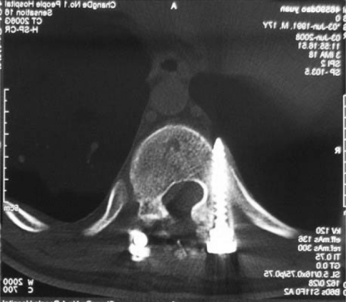
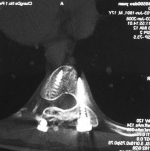
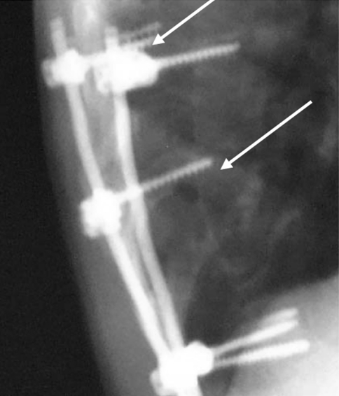
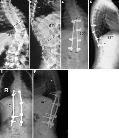
Thoracic pedicle screws have superior anchoring strength compared with other available fixation techniques. However, these are not universally accepted in many developing countries because of the concerns regarding safety and complications. In addition, there is evidence that pedicle morphology is unique in Chinese patients. The goal of this study was to analyze the complications seen at our institution, while using thoracic pedicle screws for the treatment of thoracic deformity, and to determine the safety of our techniques for the treatment of thoracic deformity in a Chinese population. From 1998 to 2005, there were 208 thoracic deformity patients treated at our institution, 70 of whom were male and 138 were female. Their age ranged from 11 to 55 years (mean of 14.9 years). All of them underwent corrective deformity surgery using posterior pedicle screw systems and follow-up was available for at least 3 years. Etiologic diagnoses included adolescent idiopathic scoliosis in 119 patients, congenital kyphoscoliosis in 38, adult scoliosis in 37 and undetermined in 14. Screw positions were evaluated using intraoperative and postoperative radiographs and a CT scan was performed when a concern for screw malposition was present. All radiographic evaluations were carried out in a double-blinded fashion. A total of 1,123 thoracic pedicle screws were inserted (5.4 thoracic screws/patient). The deformity correction rate was 81, 65 and 62% for idiopathic, congenital and adult scoliosis patients, respectively. The overall complication rate was 16.5% at the final follow-up. Complication rates directly and indirectly related to pedicle screws were 7.2 and 9.3%, respectively. There were no significant screw-related neurologic or visceral complications that adversely affected long-term results. The complications seen with thoracic pedicle screws in a Chinese population were similar to other populations and could be utilized safely for the treatment of thoracic deformity in this population.


Figures

Fig. 1
Fig. 1
Pedicle screw placement in the thoracic spine. Note the right screw tip breached the cortical bone of the thoracic vertebrae anteriorly. As the screw tip is far away from the vascular structures, it can be left in place
Fig. 2
Fig. 2
Postoperative CT scan demonstrating medial placement of a left thoracic pedicle screw. This patient experienced postoperative radicular pain and required revision surgery for screw removal
Fig. 3
Fig. 3
Post-operative X-ray shows rostral aberrant placement of a screw (lower arrow) and loss of fixation and screw pull out of a thoracic screw (upper arrow)
Fig. 4
Fig. 4
An 11-year- old boy with a congenital hemivertebra at T11. The angulation of the coronal and sagittal plane was 42 and 51, respectively. The sagittal view shows a hyperkyphosis at the thoracolumbar junctional region (a and b). Postoperative 3 months follow-up radiographs demonstrated that the angulation of the coronal and sagittal views was corrected to 20 and 38, respectively. However, the anterioposterior and lateral view (c and d) shows loosening and pullout of two screws and this patient underwent revision surgery. Four-year follow-up image (e and f). The shoulder and pelvis achieved good balance during follow-up

References

    1. Kuklo TR, Lenke LG, O’Brien MF, Lehman RA, Jr, Polly DW, Jr, Schroeder TM. Accuracy and efficacy of thoracic pedicle screws in curves more than 90 degrees. Spine. 2005;30:222–226. doi: 10.1097/01.brs.0000150482.26918.d8. - DOI - PubMed
    1. Hou S, Hu R, Shi Y. Pedicle morphology of the lower thoracic and lumbar spine in a Chinese population. Spine. 1993;18:1850–1855. doi: 10.1097/00007632-199310000-00021. - DOI - PubMed
    1. Liljenqvist U, Lepsien U, Hackenberg L, Niemeyer T, Halm H. Comparative analysis of pedicle screw and hook instrumentation in posterior correction and fusion of idiopathic thoracic scoliosis. Eur Spine J. 2002;11:336–343. doi: 10.1007/s00586-002-0415-9. - DOI - PMC - PubMed
    1. Liljenqvist UR, Halm HF, Link TM. Pedicle screw instrumentation of the thoracic spine in idiopathic scoliosis. Spine. 1997;22:2239–2245. doi: 10.1097/00007632-199710010-00008. - DOI - PubMed
    1. Ruofu Z, Huilin Y, Xiaoyun H, et al. CT evaluation of cervical pedicle in a Chinese population for surgical application of transpedicular screw placement. Surg Radiol Anat. 2008;30:389–396. doi: 10.1007/s00276-008-0339-0. - DOI - PubMed