

Acute demyelinating encephalomyelitis after anti-venom therapy in Russell's viper bite
- PMID: 20237970
- PMCID: PMC3550494
- DOI: 10.1007/s13181-010-0015-8
Acute demyelinating encephalomyelitis after anti-venom therapy in Russell's viper bite
Abstract
Introduction: Russell's viper is a commonly encountered venomous snake in India. Morbidity and mortality following envenomation and the treatment thereof are frequent. We report a rarely seen complication after a treated Russell's viper bite.
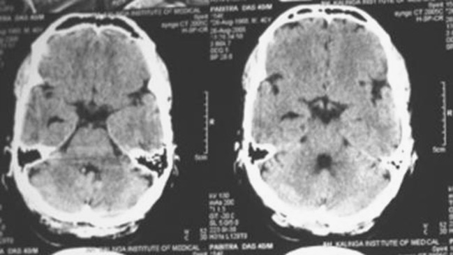
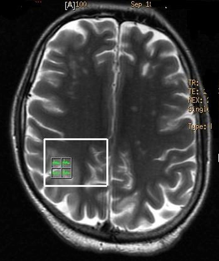
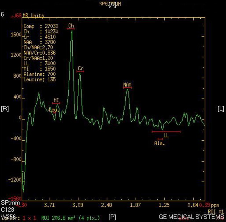
Case report: A 36-year-old male farmer received 30 vials polyvalent anti-snake venom after a viper bite to his right leg. Improvement in initial hematemesis and circulatory shock was followed by acute renal failure managed with regular hemodialysis. He displayed no abnormalities on neurological examination at admission. Fourth day onwards his neurologic status started deteriorating with development of behavioral abnormalities, hemi-spatial neglect of left upper limb, paralysis of left facial nerve, left upper limb, and right lower limb. Acute disseminated encephalomyelitis was confirmed on magnetic resonance imaging (MRI) of brain with typical spectroscopic characteristics. High dose methyl prednisolone was administered and a rapid recovery followed.
Conclusion: Russell's viper bite followed by treatment with antivenom may be complicated by the development of immune complex mediated demyelination and development of acute disseminated encephalomyelitis. MRI spectroscopy helps in early identification of demyelination and in a definite diagnosis. Treatment with corticosteroids was associated with resolution of symptoms in this case.
Figures
Similar articles
-
Neutrophil-mediated erythrophagocytosis following Russell's viper (Daboia russelii) bite.Toxicon. 2023 Jun 1;228:107111. doi: 10.1016/j.toxicon.2023.107111. Epub 2023 Apr 13. Toxicon. 2023. PMID: 37060927
-
Neurotoxicity in Russell's viper (Daboia russelii) envenoming in Sri Lanka: a clinical and neurophysiological study.Clin Toxicol (Phila). 2016 Jun;54(5):411-9. doi: 10.3109/15563650.2016.1143556. Epub 2016 Feb 29. Clin Toxicol (Phila). 2016. PMID: 26923566
-
Russell's viper envenomation induces rectus sheath haematoma.Toxicon. 2023 Mar 1;224:107037. doi: 10.1016/j.toxicon.2023.107037. Epub 2023 Jan 21. Toxicon. 2023. PMID: 36690089
-
Hypopituitarism following envenoming by Russell's vipers (Daboia siamensis and D. russelii) resembling Sheehan's syndrome: first case report from Sri Lanka, a review of the literature and recommendations for endocrine management.QJM. 2011 Feb;104(2):97-108. doi: 10.1093/qjmed/hcq214. Epub 2010 Nov 28. QJM. 2011. PMID: 21115460 Review.
-
Proteomic analysis reveals geographic variation in venom composition of Russell's Viper in the Indian subcontinent: implications for clinical manifestations post-envenomation and antivenom treatment.Expert Rev Proteomics. 2018 Oct;15(10):837-849. doi: 10.1080/14789450.2018.1528150. Expert Rev Proteomics. 2018. PMID: 30247947 Review.
Cited by
-
Identifying the snake: First scoping review on practices of communities and healthcare providers confronted with snakebite across the world.PLoS One. 2020 Mar 5;15(3):e0229989. doi: 10.1371/journal.pone.0229989. eCollection 2020. PLoS One. 2020. PMID: 32134964 Free PMC article.
-
Cerebral Complications of Snakebite Envenoming: Case Studies.Toxins (Basel). 2022 Jun 27;14(7):436. doi: 10.3390/toxins14070436. Toxins (Basel). 2022. PMID: 35878174 Free PMC article. Review.
-
Acute Reversible Ischemic Stroke after Snake Bite.Indian J Crit Care Med. 2018 Aug;22(8):611-612. doi: 10.4103/ijccm.IJCCM_455_17. Indian J Crit Care Med. 2018. PMID: 30186014 Free PMC article.
-
Reversible posterior leukoencephalopathy in a venomous snake (Bothrops asper) bite victim.Am J Trop Med Hyg. 2012 Mar;86(3):496-8. doi: 10.4269/ajtmh.2012.11-0610. Am J Trop Med Hyg. 2012. PMID: 22403325 Free PMC article.
-
Parkinsonism Associated with Snakebite.Ann Afr Med. 2024 Jul 1;23(3):518-522. doi: 10.4103/aam.aam_151_23. Epub 2024 Jul 20. Ann Afr Med. 2024. PMID: 39034585 Free PMC article.
References
Publication types
MeSH terms
Substances
LinkOut - more resources
Full Text Sources
