

Pyostomatitis vegetans: cellular immune profile and expression of IL-6, IL-8 and TNF-alpha
- PMID: 20237982
- PMCID: PMC2825530
- DOI: 10.1007/s12105-009-0149-7
Pyostomatitis vegetans: cellular immune profile and expression of IL-6, IL-8 and TNF-alpha
Abstract
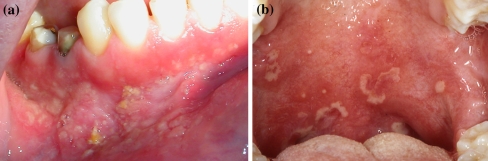
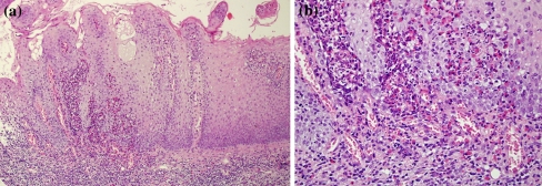
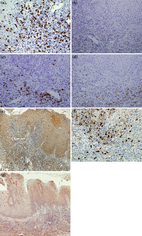
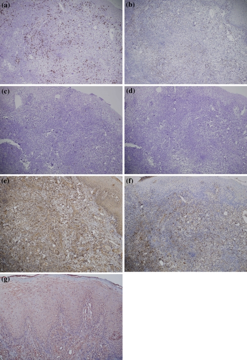
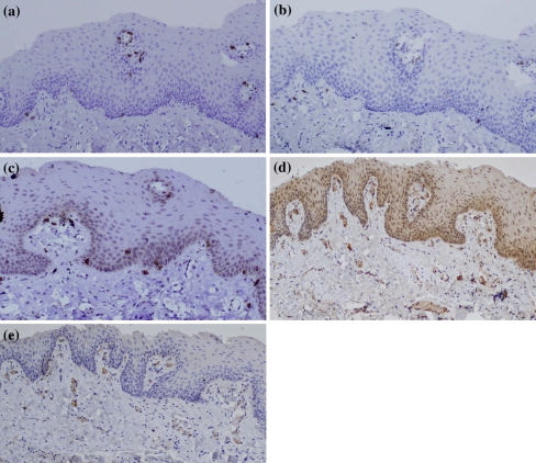
The aim of this study was to investigate the cellular immune profile and the expression of IL-6, IL-8 and TNF-alpha in tissue biopsies of pyostomatitis vegetans (PV). Working hypothesis was that knowledge of the cellular immune profile and role of mediators such as IL-6, IL-8 AND TNF-alpha may contribute to a better understanding of the pathogenesis of this rare entity. Archival tissues from three patients with clinically and histologically confirmed PV were studied. Analysis of the immune profile of the cellular infiltrate and expression of IL-6 and IL-8 were evaluated by immunohistochemistry. ISH was performed to evaluate the expression of TNF-alpha. Biopsy tissues from erythema multiforme, recurrent aphthous stomatitis, lichen planus and normal buccal mucosa were analyzed as controls. All patients were affected by multiple mucosal ulcerations and yellow pustules mainly located in the vestibular, gingival and palatal mucosa. Histopathologically, all specimens showed ulcerated epithelium with characteristic intraepithelial and/or subepithelial microabscesses containing abundant eosinophils plus a mixed infiltrate composed of lymphocytes and neutrophils. Cellular immune profile of the inflammatory infiltrate revealed a predominance of T-lymphocytes, mainly of cytotoxic (CD3+/CD8+) phenotype, over B-cells. CD20+ B-lymphocytes were also identified to a lesser degree among the lymphoid cells present in the lamina propria. Overexpression of IL-6 and TNF-alpha was found in both epithelial and inflammatory mononuclear cells. IL-8 expression was shown in the mononuclear cells scattered among the inflammatory infiltrate. Similar findings of overexpression of IL-6, IL-8 and TNF-alpha were, however, found in control tissues. In PV lesions, the inflammatory infiltrate shows a predominance of cytotoxic lymphocytes. Expression of IL-6, IL-8 and TNF-alpha, although not specific to PV, appears up-regulated thus these cytokines would represent a suitable therapeutic target. However, the complexity of the cytokine network and their numerous functions require further studies in order to confirm our findings.
Figures
Similar articles
-
Immunolocalization of tumor necrosis factor-alpha expressing cells in recurrent aphthous ulcer lesions (RAU).J Oral Pathol Med. 2000 Jan;29(1):19-25. doi: 10.1034/j.1600-0714.2000.290104.x. J Oral Pathol Med. 2000. PMID: 10678712
-
Polymorphonuclear neutrophils and their mediators in gingival tissues from generalized aggressive periodontitis.J Periodontol. 2001 Nov;72(11):1545-53. doi: 10.1902/jop.2001.72.11.1545. J Periodontol. 2001. PMID: 11759866
-
Immunohistochemical characterization of oral mucosal lesions in cats with chronic gingivostomatitis.J Comp Pathol. 2011 May;144(4):239-50. doi: 10.1016/j.jcpa.2010.09.173. Epub 2010 Nov 16. J Comp Pathol. 2011. PMID: 21084098
-
Pyostomatitis vegetans.Clin Exp Dermatol. 2004 Jan;29(1):1-7. doi: 10.1111/j.1365-2230.2004.01438.x. Clin Exp Dermatol. 2004. PMID: 14723710 Review.
-
Pyostomatitis vegetans. Report of a case and review of the literature.J Am Acad Dermatol. 1989 Aug;21(2 Pt 2):381-7. J Am Acad Dermatol. 1989. PMID: 2666469 Review.
Cited by
-
Skin manifestations of inflammatory bowel disease.Front Physiol. 2012 Feb 6;3:13. doi: 10.3389/fphys.2012.00013. eCollection 2012. Front Physiol. 2012. PMID: 22347192 Free PMC article.
-
Unravelling the Oral-Gut Axis: Interconnection Between Periodontitis and Inflammatory Bowel Disease, Current Challenges, and Future Perspective.J Crohns Colitis. 2024 Aug 14;18(8):1319-1341. doi: 10.1093/ecco-jcc/jjae028. J Crohns Colitis. 2024. PMID: 38417137 Free PMC article. Review.
-
Characterization of Cytokines and Proliferation Marker Ki67 in Cleft Affected Lip Tissue.Medicina (Kaunas). 2019 Aug 22;55(9):518. doi: 10.3390/medicina55090518. Medicina (Kaunas). 2019. PMID: 31443525 Free PMC article.
-
Pyodermatitis-Pyostomatitis Vegetans: The Role of Langerin Deficiency in Disease Pathogenesis.J Clin Med. 2025 Jun 12;14(12):4198. doi: 10.3390/jcm14124198. J Clin Med. 2025. PMID: 40565944 Free PMC article.
-
Oral manifestations serve as potential signs of ulcerative colitis: A review.Front Immunol. 2022 Sep 29;13:1013900. doi: 10.3389/fimmu.2022.1013900. eCollection 2022. Front Immunol. 2022. PMID: 36248861 Free PMC article. Review.
References
-
- Nigen S, Poulin Y, Rochette L, et al. Pyodermatitis-pyostomatitis vegetans: two cases and a review of the literature. J Cutan Med Surg. 2003;7:250–255. - PubMed
MeSH terms
Substances
LinkOut - more resources
Full Text Sources
Research Materials
