Review
. 2010 Mar;4(1):84-93.
doi: 10.1007/s12105-009-0158-6. Epub 2010 Jan 26.

The small round blue cell tumors of the sinonasal area

Affiliations
Review

The small round blue cell tumors of the sinonasal area

Julia A Bridge et al. Head Neck Pathol. 2010 Mar.

Abstract

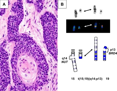
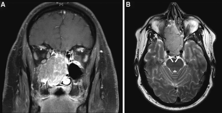
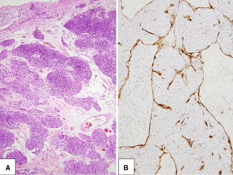
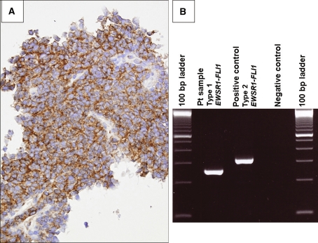
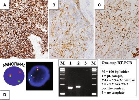
The diagnostic classification of small round blue cell tumors of the sinonasal area to include diverse malignancies of epithelial, hematolymphoid, neuroectodermal, and mesenchymal origin is challenging to the surgical pathologist using conventional histopathologic approaches because the cytomorphologic features are often overlapping or indistinctive. Rare or occasional clinical presentations in atypical age groups or unusual locations, as well as small biopsy samples may further complicate the differential diagnosis. Immunohistochemistry represents an extensively investigated ancillary technique that may aid in the provision of a definitive diagnosis. In recent years, certain small round blue cell tumors have been shown by cytogenetic analysis to have specific and primary chromosomal alterations, providing clinicians with a valuable tool to enhance their diagnostic armamentarium. The addition of molecular cytogenetic [fluorescence in situ hybridization (FISH), comparative genomic hybridization (CGH)] and molecular pathologic [polymerase chain reaction (PCR) and reverse transcriptase (RT)-PCR] approaches has further enhanced the sensitivity and accuracy of detecting these genetic alterations including assessment in formalin-fixed, paraffin-embedded tissues. Establishing an accurate diagnosis of a small round blue cell tumor of the sinonasal tract frequently requires adjunctive studies including immunohistochemical and molecular analyses.


Figures

Fig. 1
Fig. 1
aNUT-rearranged midline carcinoma; monomorphic, poorly differentiated carcinoma with evidence of squamous differentiation. This neoplasm can mimic other undifferentiated neoplasms such as squamous cell carcinoma, Ewing’s sarcoma, and lymphoma. This particular patient initially received an erroneous diagnosis of squamous cell carcinoma. b Partial G-banded karyotype, FISH conducted with a custom-designed BRD4-NUT dual fusion probe set, and schematic illustrating the t(15;19)(q14;p13) identified in a subset of NUT-rearranged midline carcinomas
Fig. 2
Fig. 2
a and b Coronal and axial magnetic resonance images demonstrating a large SNUC mass filling both sides of the nasal cavity, bilateral ethmoid sinuses, and the right maxillary sinus in a 41-year-old man who presented with a 6-month history of nasal obstruction unresponsive to decongestants, steroids, or antibiotics. Arrows (part b) indicate carotid arteries with adjacent tumor involvement
Fig. 3
Fig. 3
a Common to all grades of olfactory neuroblastoma is the lobular growth pattern. b Characteristic S100-positive sustentacular cells
Fig. 4
Fig. 4
a Characteristic membranous CD99 immunoreactivity in a maxillary sinus extraskeletal Ewing’s sarcoma. b The pathognomonic EWSR1-FLI1 fusion transcript identified by RT-PCR analysis
Fig. 5
Fig. 5
a CD56 immunostaining in a solid variant ARMS of the ethmoid sinus. b and c Synaptophysin and cytokeratin immunostaining, respectively in an ARMS of the nasopharynx. dSplit red and green signals indicative of a FOXO1 rearrangement characteristic of ARMS are identified by interphase FISH analysis with a FOXO1 breakapart probe (bottom left). PAX7-FOXO1 and PAX3-FOXO1 fusion transcripts identified by RT-PCR analysis (bottom right)

Similar articles

Cited by

References

    1. Jemal A, Siegel R, Ward E, et al. Cancer statistics. CA Cancer J Clin. 2009;59:225–249. doi: 10.3322/caac.20006. - DOI - PubMed
    1. Barnes L, Everson JW, Reichart P, Sidransky D (eds) (2005) Pathology and genetics of head and neck tumours. Kleihues P, Sobin LH, series eds. World health organization classification of tumours. Lyon, France: IARC Press.
    1. Franchi A, Moroni M, Massi D, et al. Sinonasal undifferentiated carcinoma, nasopharyngeal-type undifferentiated carcinoma, and keratinizing and nonkeratinizing squamous cell carcinoma express different cytokeratin patterns. Am J Surg Pathol. 2002;26:1597–1604. doi: 10.1097/00000478-200212000-00007. - DOI - PubMed
    1. Cerelli LA, Holst VA, Brandwein MS, et al. Sinonasal undifferentiated carcinoma: Immunohistochemical profile and lack of EBV association. Am J Surg Pathol. 2001;25:156–163. doi: 10.1097/00000478-200102000-00003. - DOI - PubMed
    1. French CA, Kutok JL, Faquin WC, et al. Midline carcinoma of children and young adults with NUT rearrangement. J Clin Oncol. 2004;22:4135–4139. doi: 10.1200/JCO.2004.02.107. - DOI - PubMed

MeSH terms

LinkOut - more resources