

Photoreceptor sensory cilia and inherited retinal degeneration
- PMID: 20238021
- PMCID: PMC2888132
- DOI: 10.1007/978-1-4419-1399-9_26
Photoreceptor sensory cilia and inherited retinal degeneration
Abstract
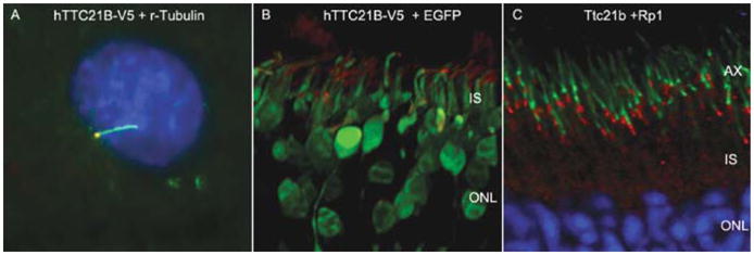
The outer segments of photoreceptor cells are specialized sensory cilia, and share many features with other primary and sensory cilia. Like other cilia, photoreceptor sensory cilium (PSC) comprises a membrane domain of outer segment and its cytoskeleton. We have recently identified the protein components of mouse PSCs, and found that the list of PSC proteins, called the PSC proteome, contains many novel cilia proteins. Studies have shown that many of the identified retinal degeneration disease genes encode proteins which are part of the PSC. Furthermore, mutations in genes encoding proteins expressed both in photoreceptors and other cilia result in systemic diseases, such as Usher syndrome, Bardet-Biedl syndrome (BBS), and Senior-Loken syndrome that involve retinal degeneration along with other disorders consequent to cilia dysfunction such as deafness and polycystic kidney disease. Based on these findings, we hypothesize that genes that encode proteins required for formation of PSCs are good candidate retinal degeneration disease genes. This chapter will summarize our studies on identifying novel PSC proteins from the PSC proteome. As an example of these studies, we demonstrated that tetratricopeptide the repeat domain 21B (TTC21B) protein is a novel PSC protein and is required for normal cilia formation in primary and photoreceptor sensory cilia.
Figures
Similar articles
-
The proteome of the mouse photoreceptor sensory cilium complex.Mol Cell Proteomics. 2007 Aug;6(8):1299-317. doi: 10.1074/mcp.M700054-MCP200. Epub 2007 May 9. Mol Cell Proteomics. 2007. PMID: 17494944 Free PMC article.
-
Accumulation of non-outer segment proteins in the outer segment underlies photoreceptor degeneration in Bardet-Biedl syndrome.Proc Natl Acad Sci U S A. 2015 Aug 11;112(32):E4400-9. doi: 10.1073/pnas.1510111112. Epub 2015 Jul 27. Proc Natl Acad Sci U S A. 2015. PMID: 26216965 Free PMC article.
-
RPGR-containing protein complexes in syndromic and non-syndromic retinal degeneration due to ciliary dysfunction.J Genet. 2009 Dec;88(4):399-407. doi: 10.1007/s12041-009-0061-7. J Genet. 2009. PMID: 20090203 Free PMC article. Review.
-
Retinal Degeneration Animal Models in Bardet-Biedl Syndrome and Related Ciliopathies.Cold Spring Harb Perspect Med. 2023 Jan 3;13(1):a041303. doi: 10.1101/cshperspect.a041303. Cold Spring Harb Perspect Med. 2023. PMID: 36596648 Free PMC article. Review.
-
Depletion of BBS Protein LZTFL1 Affects Growth and Causes Retinal Degeneration in Mice.J Genet Genomics. 2016 Jun 20;43(6):381-91. doi: 10.1016/j.jgg.2015.11.006. Epub 2016 May 5. J Genet Genomics. 2016. PMID: 27312011 Free PMC article.
Cited by
-
RNA-Seq: Improving Our Understanding of Retinal Biology and Disease.Cold Spring Harb Perspect Med. 2015 Feb 26;5(9):a017152. doi: 10.1101/cshperspect.a017152. Cold Spring Harb Perspect Med. 2015. PMID: 25722474 Free PMC article. Review.
-
Transcriptional program of ciliated epithelial cells reveals new cilium and centrosome components and links to human disease.PLoS One. 2012;7(12):e52166. doi: 10.1371/journal.pone.0052166. Epub 2012 Dec 31. PLoS One. 2012. PMID: 23300604 Free PMC article.
-
Gene expression changes in the retina following subretinal injection of human neural progenitor cells into a rodent model for retinal degeneration.Mol Vis. 2016 May 16;22:472-90. eCollection 2016. Mol Vis. 2016. PMID: 27217715 Free PMC article.
-
Kinesin family 17 (osmotic avoidance abnormal-3) is dispensable for photoreceptor morphology and function.FASEB J. 2015 Dec;29(12):4866-80. doi: 10.1096/fj.15-275677. Epub 2015 Jul 30. FASEB J. 2015. PMID: 26229057 Free PMC article.
-
New interaction partners for Nek4.1 and Nek4.2 isoforms: from the DNA damage response to RNA splicing.Proteome Sci. 2015 Feb 26;13:11. doi: 10.1186/s12953-015-0065-6. eCollection 2015. Proteome Sci. 2015. PMID: 25798074 Free PMC article.
References
-
- Allen RA. Isolated cilia in inner retinal neurons and in retinal pigment epithelium. J Ultrastruct Res. 1965;12:730–747. - PubMed
-
- Ansley SJ, Badano JL, Blacque OE, et al. Basal body dysfunction is a likely cause of pleiotropic Bardet-Biedl syndrome. Nature. 2003;425:628–633. - PubMed
-
- Badano JL, Mitsuma N, Beales PL, et al. The ciliopathies: an emerging class of human genetic disorders. Annu Rev Genomics Hum Genet. 2006;7:125–148. - PubMed
Publication types
MeSH terms
Substances
Grants and funding
LinkOut - more resources
Full Text Sources
Other Literature Sources
Medical
Research Materials
