

Gene therapy in the Retinal Degeneration Slow model of retinitis pigmentosa
- PMID: 20238065
- PMCID: PMC3161507
- DOI: 10.1007/978-1-4419-1399-9_70
Gene therapy in the Retinal Degeneration Slow model of retinitis pigmentosa
Abstract
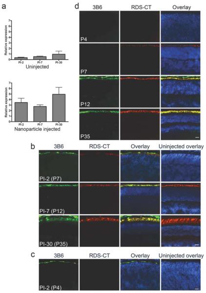
Human blinding disorders are often initiated by hereditary mutations that insult rod and/or cone photoreceptors and cause subsequent cellular death. Generally, the disease phenotype can be predicted from the specific mutation as many photoreceptor genes are specific to rods or cones; however certain genes, such as Retinal Degeneration Slow (RDS), are expressed in both cell types and cause different forms of retinal disease affecting rods, cones, or both photoreceptors. RDS is a transmembrane glycoprotein critical for photoreceptor outer segment disc morphogenesis, structural maintenance, and renewal. Studies using animal models with Rds mutations provide valuable insight into Rds gene function and regulation; and a better understanding of the physiology, pathology, and underlying degenerative mechanisms of inherited retinal disease. Furthermore, these models are an excellent tool in the process of developing therapeutic interventions for the treatment of inherited retinal degenerations. In this paper, we review these topics with particular focus on the use of rds models in gene therapy.
Figures
Similar articles
-
Gene delivery to mitotic and postmitotic photoreceptors via compacted DNA nanoparticles results in improved phenotype in a mouse model of retinitis pigmentosa.FASEB J. 2010 Apr;24(4):1178-91. doi: 10.1096/fj.09-139147. Epub 2009 Dec 1. FASEB J. 2010. PMID: 19952284 Free PMC article.
-
Mutations and polymorphisms in the human peripherin-RDS gene and their involvement in inherited retinal degeneration.Hum Mutat. 1996;8(4):297-303. doi: 10.1002/(SICI)1098-1004(1996)8:4<297::AID-HUMU1>3.0.CO;2-5. Hum Mutat. 1996. PMID: 8956033
-
Expression of Bcl-2 protects against photoreceptor degeneration in retinal degeneration slow (rds) mice.J Neurosci. 2000 Mar 15;20(6):2150-4. doi: 10.1523/JNEUROSCI.20-06-02150.2000. J Neurosci. 2000. PMID: 10704489 Free PMC article.
-
The spectrum of retinal dystrophies caused by mutations in the peripherin/RDS gene.Prog Retin Eye Res. 2008 Mar;27(2):213-35. doi: 10.1016/j.preteyeres.2008.01.002. Epub 2008 Jan 26. Prog Retin Eye Res. 2008. PMID: 18328765 Review.
-
The role of Rds in outer segment morphogenesis and human retinal disease.Ophthalmic Genet. 2006 Dec;27(4):117-22. doi: 10.1080/13816810600976806. Ophthalmic Genet. 2006. PMID: 17148038 Review.
Cited by
-
Direct gene transfer with compacted DNA nanoparticles in retinal pigment epithelial cells: expression, repeat delivery and lack of toxicity.Nanomedicine (Lond). 2012 Apr;7(4):521-39. doi: 10.2217/nnm.11.158. Epub 2012 Feb 23. Nanomedicine (Lond). 2012. PMID: 22356602 Free PMC article.
-
Gene delivery to the retina: from mouse to man.Methods Enzymol. 2012;507:255-74. doi: 10.1016/B978-0-12-386509-0.00013-2. Methods Enzymol. 2012. PMID: 22365778 Free PMC article.
-
Clinical and Rehabilitative Management of Retinitis Pigmentosa: Up-to-Date.Curr Genomics. 2011 Jun;12(4):250-9. doi: 10.2174/138920211795860125. Curr Genomics. 2011. PMID: 22131870 Free PMC article.
References
-
- Acland GM, Aguirre GD, Ray J, et al. Gene therapy restores vision in a canine model of childhood blindness. Nat Genet. 2001;28:92–95. - PubMed
-
- Ali RR, Sarra GM, Stephens C, et al. Restoration of photoreceptor ultrastructure and function in retinal degeneration slow mice by gene therapy. Nat Genet. 2000;25:306–310. - PubMed
-
- Allen D, Kenna PF, Palfi A, et al. Development of strategies for conditional RNA interference. J Gene Med. 2007;9:287–298. - PubMed
Publication types
MeSH terms
Substances
Grants and funding
LinkOut - more resources
Full Text Sources
Medical
