Review
. 2010 Apr;38(2):384-7.
doi: 10.1042/BST0380384.

Orchestrating serine resolvases

Affiliations
Review

Orchestrating serine resolvases

Phoebe A Rice et al. Biochem Soc Trans. 2010 Apr.

Abstract

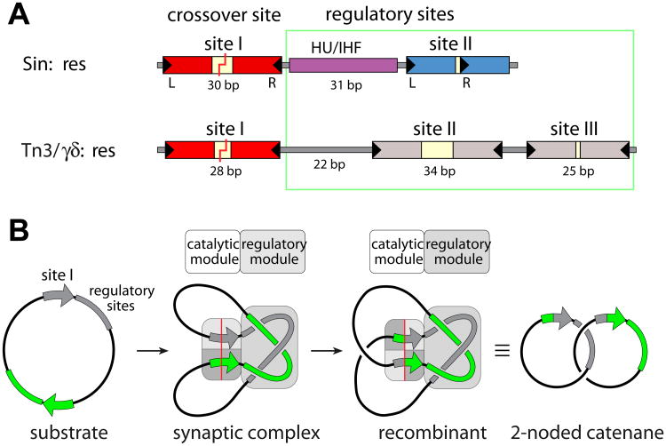
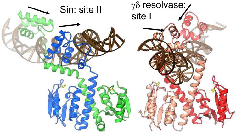
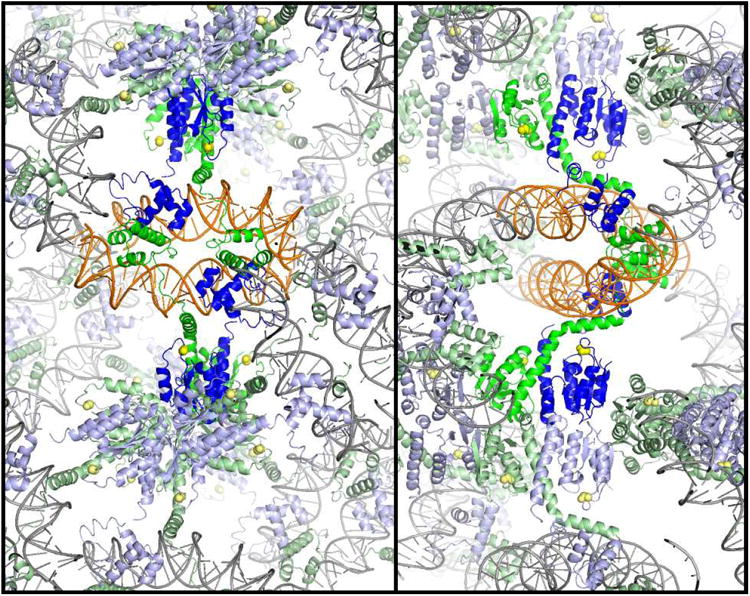
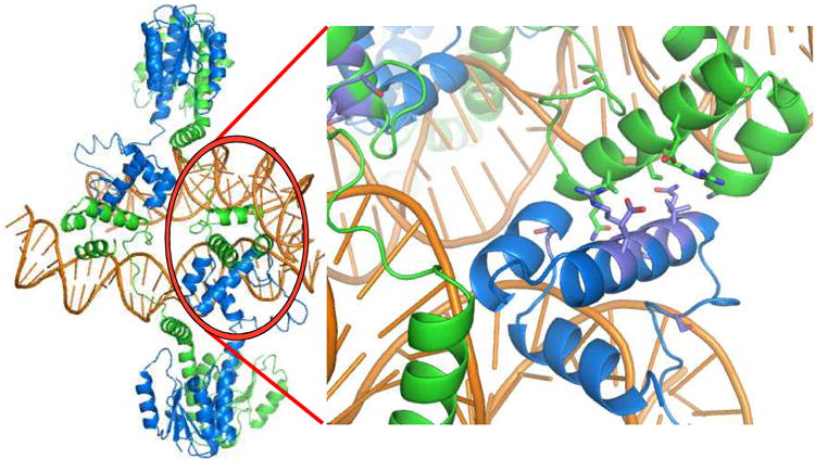
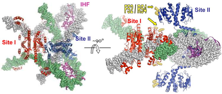
A remarkable feature of the serine resolvases is their regulation: the wild-type enzymes will catalyse intra- but not inter-molecular recombination, can sense the relative orientation of their sites and can exchange strands directionally, despite the fact that there is no net release of chemical bond energy. The key to this regulation is that they are only active within a large intertwined complex called the 'synaptosome'. Because substrate topology greatly facilitates (or, in other cases, inhibits) formation of the synaptosome, it acts as a 'topological filter'. Within the defined topology of the synaptosome, strand exchange releases supercoiling tension, providing an energy source to bias the reaction direction. The regulatory portion of this complex contains additional copies of the recombinase and sometimes other DNA-bending proteins. We are using a combination of X-ray crystallography, biochemistry and genetics to model the full synaptic complex and to understand how the regulatory portion activates the crossover-site-bound recombinases.


Figures

Figure 1
Figure 1. Recombination sites and synaptosome cartoons
a) Comparison of the full recombination sites for Sin (top) and γδ/Tn3 resolvases (bottom). The Sin site binds two Sin dimers (at sites I and II) and a DNA bending protein (HU or IHF), while the γδ/Tn3 site binds three recombinase dimers. DNA breakage and rejoining occurs Site I. For Sin, the individual subsites are colored to match the proteins bound to them in figure 3. b) Cartoon of resolution. Synaptosome formation brings two site Is together and traps 3 interdomainal supercoils. A 180° rotation within the site I-bound tetramer creates recombinant products, which are catenated daughter circles. Adapted from [16].
Figure 2
Figure 2. Building a structural model of the Sin synaptosome
a) Structure of the Sin – site II complex (left), and comparison to the WT γδ resolvase – site I complex (right) [5, 16]. Arrows show the directions of individual half site sequences, which form a direct repeat for Sin site II and an inverted repeat for site I. Active site serines are shown as yellow balls and sticks. b) Packing of the Sin – Site II complex. Two orthogonal cross-sections of the crystal lattice are shown, viewed down the z axis. Individual Sin dimer – site II complexes are colored as in figure 1a, with two dimers representing a biologically relevant tetramer shown in brighter colors. The crystal contacts include end-to-end stacking of the DNA duplexes, contacts between DNA binding domains, and contacts between catalytic domains. c) The biologically relevant tetramer (left) is found within the crystal, and is mediated by contacts between DNA binding domains. Residues whose mutation interferes with site II – site II contacts and recombination in vivo are shown in the closeup at right.
Figure 3
Figure 3
Model of the Sin synaptosome. Four structures were docked together to create this model: the activated γδ resolvase tetramer - site I complex (red proteins), the IHF-DNA complex (magenta proteins), the Sin – site II complex (blue proteins), and one turn of model-built B-form DNA to continue the path of the DNA past the end of site II [16]. One DNA partner is in gray, the other green. Note that the protein colors correspond to those of their sites in figure 1a.

Similar articles

Cited by

References

    1. Grindley ND, Whiteson KL, Rice PA. Mechanisms of Site-Specific Recombination. Annu Rev Biochem. 2006;75:567–605. - PubMed
    1. Rowland SJ, Dyke KG. Characterization of the staphylococcal beta-lactamase transposon Tn552. Embo J. 1989;8:2761–2773. - PMC - PubMed
    1. Highlander SK, Hulten KG, Qin X, Jiang H, Yerrapragada S, Mason EO, Jr, Shang Y, Williams TM, Fortunov RM, Liu Y, et al. Subtle genetic changes enhance virulence of methicillin resistant and sensitive Staphylococcus aureus. BMC Microbiol. 2007;7:99. - PMC - PubMed
    1. Li W, Kamtekar S, Xiong Y, Sarkis GJ, Grindley ND, Steitz TA. Structure of a synaptic gammadelta resolvase tetramer covalently linked to two cleaved DNAs. Science. 2005;309:1210–1215. - PubMed
    1. Yang W, Steitz TA. Crystal structure of the site-specific recombinase gamma delta resolvase complexed with a 34 bp cleavage site. Cell. 1995;82:193–207. - PubMed

Publication types

MeSH terms

LinkOut - more resources