. 2010;12(2):R44.
doi: 10.1186/ar2954. Epub 2010 Mar 18.

Development of proteoglycan-induced arthritis depends on T cell-supported autoantibody production, but does not involve significant influx of T cells into the joints

Affiliations

Development of proteoglycan-induced arthritis depends on T cell-supported autoantibody production, but does not involve significant influx of T cells into the joints

Adrienn Angyal et al. Arthritis Res Ther. 2010.

Abstract

Introduction: Inflammatory joint destruction in rheumatoid arthritis (RA) may be triggered by autoantibodies, the production of which is supported by autoreactive T cells. Studies on RA and animal models of the disease suggest that T cells recruited in the joints can locally initiate or propagate arthritis. Herein, we investigated the role of joint-homing versus lymphoid organ-homing T cells in the development of proteoglycan-induced arthritis (PGIA), an autoimmune model of RA.

Methods: To identify T cells migrating to the joints before and during development of autoimmune arthritis, we transferred fluorescence-labeled T cells, along with antigen-presenting cells, from BALB/c mice with PGIA to naïve syngeneic severe combined immunodeficient (SCID) mice. We then monitored the recruitment of donor T cells in the ankle joints and joint-draining lymph nodes of the recipients using in vivo two-photon microscopy and ex vivo detection methods. To limit T-cell access to the joints, we selectively depleted T cells in the blood circulation by treatment with FTY720, an inhibitor of lymphocyte egress from lymphoid organs. Reduction of T cell presence in both lymphoid organs and blood was achieved by injection of donor cells from which T cells were removed prior to transfer. T and B cells were quantitated by flow cytometry, and antigen (PG)-specific responses were assessed by cell proliferation and serum antibody assays.

Results: Despite development of adoptively transferred arthritis in the recipient SCID mice, we found very few donor T cells in their joints after cell transfer. Treatment of recipient mice with FTY720 left the T-cell pool in the lymphoid organs intact, but reduced T cells in both peripheral blood and joints. However, FTY720 treatment failed to inhibit PGIA development. In contrast, arthritis was not seen in recipient mice after transfer of T cell-depleted cells from arthritic donors, and serum autoantibodies to PG were not detected in this group of mice.

Conclusions: Our results suggest that antigen-specific T cells, which home to lymphoid organs and provide help to B cells for systemic autoantibody production, play a greater role in the development and progression of autoimmune arthritis than the small population of T cells that migrate to the joints.


Figures
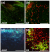
Figure 1
Figure 1
T cells or B cells, transferred from arthritic BALB/c mice to severe combined immunodeficient (SCID) mice, are detectable by in vivo imaging in the popliteal lymph nodes (LNs) but not in the joints of the recipient mice. (a) Two-photon microscopy (TPM) image of the ankle joint of a SCID mouse 2 days after transfer of CellTracker Red (CMTPX)-labeled T cells and unlabeled non-T cells (antigen-presenting cells, or APCs) from arthritic BALB/c donors. No red fluorescent cells are visible in the joint. Second harmonic generation (SHG) signals from collagen fibers (around blood vessels) are detected in the green fluorescence channel in two-color acquisition. (b) TPM image of the joint-draining (popliteal) LN from the same mouse shows numerous red fluorescent donor T cells that homed to the LN. SHG (green) signals are from collagen in the LN capsule and stroma. (c) TPM image of the inflamed ankle of a SCID mouse 12 days after transfer of CellTracker Red-labeled T cells and CellTracker Green (CMFDA)-labeled APCs (>85% B cells) from arthritic donors. Although this joint was heavily inflamed, no red fluorescent T cells (or green fluorescent B cells) were found by in vivo TPM imaging. SHG signals from collagen are detected in the blue channel in this three-color acquisition image. Endogenous (auto) fluorescence from macrophages appears in light green. (d) TPM image of the popliteal LN from the same mouse shows large numbers of red fluorescent T cells and green fluorescent non-T cells (mostly B cells), which occupy the T-cell and B-cell zones of the LN, respectively. The TPM images shown are representative samples of ankle and LN images from six SCID mice at each time point. Scale bars, 100 μm.
Figure 2
Figure 2
Immunohistochemical and flow cytometric detection of T cells and granulocytes in inflamed ankle joints of severe combined immunodeficient (SCID) mice with adoptively transferred proteoglycan-induced arthritis (PGIA). The SCID mouse used for immunohistochemistry developed arthritis 9 days after receiving unlabeled cell transfer and was re-transferred with CellTracker Red-labeled T cells and unlabeled non-T cells on day 10. In vivo two-photon microscopy imaging of the inflamed ankle on day 12 revealed no red fluorescent cells. The mouse was sacrificed, and frozen sections were prepared from the ankle after imaging. (a) No red fluorescent T cells are visible in the unstained section of the inflamed ankle. (b) Immunostaining with green fluorophore-conjugated anti-CD4 monoclonal antibody (mAb) shows no evidence of CD4+ T helper cells (no yellow or green color). (c) Anti-Gr-1 mAb against granulocytes stains numerous cells in the same joint. Scale bars, 100 μm. St, synovial tissue. (d) Flow cytometry of synovial fluid cells from the arthritic ankle joints of a SCID mouse shows a small population of T cells, nearly all of which are CD4+, in the fluid. (e) Synovial fluid from arthritic SCID ankles contains a large proportion of Gr-1hi granulocytes that also express high levels of CD11b. Flow cytometry was done using synovial fluid samples from SCID mice that developed PGIA approximately 2 weeks after transfer of unlabeled cells from arthritic donors. Flow data are representative of at least six synovial fluid samples harvested from inflamed ankle joints of SCID mice with adoptively transferred PGIA.
Figure 3
Figure 3
Effects of FTY720 treatment or depletion of T cells from the transferred donor cells on circulating T cells and arthritis development in severe combined immunodeficient (SCID) mice. SCID mice, injected with lymphocytes from arthritic donors, were subjected to treatment with placebo (open circles) or FTY720 (closed triangles), as described in Materials and Methods. A separate group of SCID mice received cells from the same arthritic donors, but T cells from the donor population were depleted prior to transfer (solid circles). (a) The proportion of T cells (CD3+) among all blood leukocytes (CD45+) was monitored by flow cytometry from between day 1 after the first cell transfer and day 42 (end of experiment). Data shown are the mean ± standard error of the mean (SEM). (n = 9 to 11 mice per group; *P < 0.01 in comparison with the placebo-treated group.) (b) The SCID mice were inspected for arthritis symptoms at 4-day intervals between days 10 and 42. Incidence of proteoglycan-induced arthritis is expressed as the percentage of arthritic animals among all SCID mice in the respective groups. (c) The degree of inflammation in each paw was scored visually at 4-day intervals. Arthritis severity is expressed as the mean ± SEM of cumulative paw scores (n = 9 to 11 mice per group; *P < 0.01 in comparison with both the placebo- and FTY720-treated groups).
Figure 4
Figure 4
Treatment of immunocompetent (wild-type) BALB/c mice with FTY720 has no effect on the development of the primary form of proteoglycan (PG)-induced arthritis. (a) Short-term treatment with placebo (open circles) or FTY720 (closed triangles) started on day 49 or 50 (1 week after the third PG immunization) and ended on day 75. (b) Long-term (prophylactic) treatment was initiated after the first immunization and ended on day 70. Data shown are the mean ± standard error of the mean of cumulative arthritis scores over time (short-term treatment, n = 10 mice per group; long-term treatment, n = 16 mice per group; the difference between placebo- and FTY720-treated groups was not significant in either case). (c) Histology of the ankle joint of a placebo-treated mouse from the long-term treatment group. (d) Histology of the ankle of an FTY720-treated mouse from the long-term treatment group. Sagittal sections of decalcified and paraffin-embedded joints were stained with hematoxylin and eosin. The degree of synovial tissue hyperplasia, leukocyte infiltration, or cartilage erosion was similar in both joints. Scale bars, 250 μm. Bo, bone (talus); Jc, joint cavity; St, synovial tissue.
Figure 5
Figure 5
Proportion of lymphoid organ-homing T cells and magnitude of antigen (Ag)-specific T-cell responses in severe combined immunodeficient (SCID) mice treated with placebo or FTY720 after receiving complete donor lymphocyte transfer (T cells along with non-T cells as Ag-presenting cells) and in SCID mice receiving T cell-depleted transfer. T-cell proportions in the (a) joint-draining lymph nodes (JDLNs) and (b) spleens were measured by flow cytometry and expressed as percentage of CD3+ of total CD45+ cells. Data shown are the mean ± standard error of the mean (SEM) (n = 9 to 11 mice per group; *P < 0.05). (c) Ag (human proteoglycan)-specific spleen cell proliferation and (d) Ag-specific production of bioactive interleukin-2 (IL-2) by the same spleen cell cultures were assayed on the basis of [3H]thymidine incorporation into splenocytes and CTLL-2 cells, respectively. Results are expressed as stimulation index and are the mean ± SEM (n = 4 to 5 mice per group; *P < 0.01). Isotope incorporation by non-stimulated cell cultures was less than 1,700 cpm per well and was comparable among the three groups of mice. PG, proteoglycan.
Figure 6
Figure 6
Concentrations of proteoglycan (PG)-specific serum antibodies and percentages of spleen B cells in severe combined immunodeficient (SCID) mice treated with placebo or FTY720 after receiving complete donor lymphocyte transfer and in SCID mice receiving T cell-depleted transfer. Serum levels of (a) human PG (hPG)-specific antibodies and (b) mouse PG (mPG)-specific autoantibodies of the IgG1 isotype were determined by enzyme-linked immunosorbent assay and expressed as milligrams per milliliter and micrograms per milliliter, respectively. N.D., not detectable. (c) The proportion of B cells in the spleens of the three groups of SCID mice was measured by flow cytometry and expressed as percentage B220+ of total CD45+ cells. Data shown are the mean + standard error of the mean (n = 9 to 11 mice per group).

Comment in

  • Arthritis: where are the T cells?
    Kamradt T, Frey O. Kamradt T, et al. Arthritis Res Ther. 2010;12(3):122. doi: 10.1186/ar3008. Epub 2010 Jun 3. Arthritis Res Ther. 2010. PMID: 20537200 Free PMC article.

References

    1. McCarty DJ. In: Arthritis and Allied Conditions: A Textbook of Rheumatology. McCarty DJ, Koopman WJ, editor. Philadelphia, London: Lea & Fabiger; 1993. Clinical picture of rheumatoid arthritis; pp. 781–809.
    1. Firestein GS. In: Kelley's Textbook of Rheumatology. Ruddy S, Harris ED, Sledge CB, Kelley WN, editor. Philadelphia, PA: W.B. Saunders Co; 2005. Rheumatoid arthritis: etiology and pathogenesis of rheumatoid arthritis; pp. 996–1045.
    1. Andersson AK, Li C, Brennan FM. Recent developments in the immunobiology of rheumatoid arthritis. Arthritis Res Ther. 2008;10:204–212. doi: 10.1186/ar2370. - DOI - PMC - PubMed
    1. Nielen MM, van Schaardenburg D, Reesink HW, Twisk JW, Stadt RJ van de, Horst-Bruinsma IE van der, de Koning MH, Habibuw MR, Dijkmans BA. Simultaneous development of acute phase response and autoantibodies in preclinical rheumatoid arthritis. Ann Rheum Dis. 2006;65:535–537. doi: 10.1136/ard.2005.040659. - DOI - PMC - PubMed
    1. Weyand CM, Goronzy JJ, Takemura S, Kurtin PJ. Cell-cell interactions in synovitis. Interactions between T cells and B cells in rheumatoid arthritis. Arthritis Res. 2000;2:457–463. doi: 10.1186/ar128. - DOI - PMC - PubMed

Publication types

MeSH terms