

Encephalomyocarditis virus infection in an Italian zoo
- PMID: 20298561
- PMCID: PMC2848215
- DOI: 10.1186/1743-422X-7-64
Encephalomyocarditis virus infection in an Italian zoo
Abstract
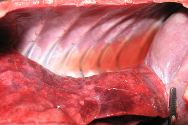
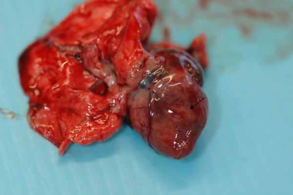
A fatal Encephalomyocarditis virus (EMCV) infection epidemic involving fifteen primates occurred between October 2006 and February 2007 at the Natura Viva Zoo. This large open-field zoo park located near Lake Garda in Northern Italy hosts one thousand animals belonging to one hundred and fifty different species, including various lemur species. This lemur collection is the most relevant and rich in Italy. A second outbreak between September and November 2008 involved three lemurs. In all cases, the clinical signs were sudden deaths generally without any evident symptoms or only with mild unspecific clinical signs. Gross pathologic changes were characterized by myocarditis (diffuse or focal pallor of the myocardium), pulmonary congestion, emphysema, oedema and thoracic fluid. The EMCV was isolated and recognized as the causative agent of both outbreaks. The first outbreak in particular was associated with a rodent plague, confirming that rats are an important risk factor for the occurrence of the EMCV infection.
Figures


References
-
- Gaskin JM, Jorge MA, Simpson CF, Lewis AL, Olson JH, Schobert EE, Wollenman EP, Marlowe C, Curtis MM. The tragedy of encephalomyocarditis virus infection in zoological parks of Florida. Proceedings American Association of Zoo Veterinarians. 1980. pp. 1–7.
-
- Gaskin JM, Andresen TL, Olsen JH, Schobert EE, Buesse D, Lynch JD, Walsh M, Citino S, Murphy D. Encephalomyocarditis in zoo animals: Recent experiences with the disease and vaccination. Proceedings of the 1st International Conference on Zoological and Avian Medicine. 1987. p. 491.
-
- McLelland D, Kirkpatrick JF, Rose K, Dixon R. Studies on encephalomyocarditis virus (EMCV) in a zoologic context. AAZV, AAWV, ARAV, NAZWV Joint Conference. 2001. p. 337.
-
- Reddacliff L, Kirkland PD, Hartley WJ, Reece RL. Encephalomyocarditis virus infections in an Australian zoo. J Zoo Wild Med. 1997;28(2):153–157. - PubMed
-
- Wells SK, Gutter AE, Soike KF, Baskin GB. Encephalomyocarditis virus: Epizootic in a zoological collection. J Zoo Wildl Med. 1989;20:291.
Publication types
MeSH terms
LinkOut - more resources
Full Text Sources
