Neuropeptidomic analysis of the embryonic Japanese quail diencephalon
- PMID: 20298575
- PMCID: PMC2851587
- DOI: 10.1186/1471-213X-10-30
Neuropeptidomic analysis of the embryonic Japanese quail diencephalon
Abstract
Background: Endogenous peptides such as neuropeptides are involved in numerous biological processes in the fully developed brain but very little is known about their role in brain development. Japanese quail is a commonly used bird model for studying sexual dimorphic brain development, especially adult male copulatory behavior in relation to manipulations of the embryonic endocrine system. This study uses a label-free liquid chromatography mass spectrometry approach to analyze the influence of age (embryonic days 12 vs 17), sex and embryonic day 3 ethinylestradiol exposure on the expression of multiple endogenous peptides in the developing diencephalon.
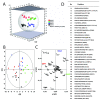
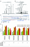
Results: We identified a total of 65 peptides whereof 38 were sufficiently present in all groups for statistical analysis. Age was the most defining variable in the data and sex had the least impact. Most identified peptides were more highly expressed in embryonic day 17. The top candidates for EE2 exposure and sex effects were neuropeptide K (downregulated by EE2 in males and females), gastrin-releasing peptide (more highly expressed in control and EE2 exposed males) and gonadotropin-inhibiting hormone related protein 2 (more highly expressed in control males and displaying interaction effects between age and sex). We also report a new potential secretogranin-2 derived neuropeptide and previously unknown phosphorylations in the C-terminal flanking protachykinin 1 neuropeptide.
Conclusions: This study is the first larger study on endogenous peptides in the developing brain and implies a previously unknown role for a number of neuropeptides in middle to late avian embryogenesis. It demonstrates the power of label-free liquid chromatography mass spectrometry to analyze the expression of multiple endogenous peptides and the potential to detect new putative peptide candidates in a developmental model.
Figures


Similar articles
-
The sexually dimorphic medial preoptic nucleus of quail: a key brain area mediating steroid action on male sexual behavior.Front Neuroendocrinol. 1996 Jan;17(1):51-125. doi: 10.1006/frne.1996.0002. Front Neuroendocrinol. 1996. PMID: 8788569 Review.
-
Impact of endocrine disrupting chemicals on reproduction in Japanese quail.Domest Anim Endocrinol. 2005 Aug;29(2):420-9. doi: 10.1016/j.domaniend.2005.02.036. Epub 2005 Apr 7. Domest Anim Endocrinol. 2005. PMID: 15998507 Review.
-
Reproductive toxicity of trenbolone acetate in embryonically exposed Japanese quail.Chemosphere. 2007 Jan;66(7):1191-6. doi: 10.1016/j.chemosphere.2006.07.085. Epub 2006 Sep 20. Chemosphere. 2007. PMID: 16989888
-
Do sex differences in the brain explain sex differences in the hormonal induction of reproductive behavior? What 25 years of research on the Japanese quail tells us.Horm Behav. 1996 Dec;30(4):627-61. doi: 10.1006/hbeh.1996.0066. Horm Behav. 1996. PMID: 9047287 Review.
-
Neuroendocrine and behavioral implications of endocrine disrupting chemicals in quail.Horm Behav. 2001 Sep;40(2):234-47. doi: 10.1006/hbeh.2001.1695. Horm Behav. 2001. PMID: 11534988 Review.
Cited by
-
Neuropeptidomics: Mass Spectrometry-Based Identification and Quantitation of Neuropeptides.Genomics Inform. 2016 Mar;14(1):12-9. doi: 10.5808/GI.2016.14.1.12. Epub 2016 Mar 31. Genomics Inform. 2016. PMID: 27103886 Free PMC article. Review.
-
Novel Single-Nucleotide Polymorphisms (SNPs) and Genetic Studies of the Shadow of Prion Protein (SPRN) in Quails.Animals (Basel). 2024 Aug 26;14(17):2481. doi: 10.3390/ani14172481. Animals (Basel). 2024. PMID: 39272266 Free PMC article.
-
Quantitation of endogenous peptides using mass spectrometry based methods.Curr Opin Chem Biol. 2013 Oct;17(5):801-8. doi: 10.1016/j.cbpa.2013.05.030. Epub 2013 Jun 18. Curr Opin Chem Biol. 2013. PMID: 23790312 Free PMC article. Review.
-
Quantitative peptidomics for discovery of circadian-related peptides from the rat suprachiasmatic nucleus.J Proteome Res. 2013 Feb 1;12(2):585-93. doi: 10.1021/pr300605p. Epub 2013 Jan 11. J Proteome Res. 2013. PMID: 23256577 Free PMC article.
-
Mass Spectrometry-based Proteomics and Peptidomics for Systems Biology and Biomarker Discovery.Front Biol (Beijing). 2012 Aug 1;7(4):313-335. doi: 10.1007/s11515-012-1218-y. Front Biol (Beijing). 2012. PMID: 24504115 Free PMC article.
References
Publication types
MeSH terms
Substances
LinkOut - more resources
Full Text Sources

