. 2010 Jun 16;168(1):300-13.
doi: 10.1016/j.neuroscience.2010.03.012. Epub 2010 Mar 15.

Neural heterogeneities and stimulus properties affect burst coding in vivo

Affiliations

Neural heterogeneities and stimulus properties affect burst coding in vivo

O Avila-Akerberg et al. Neuroscience. .

Abstract

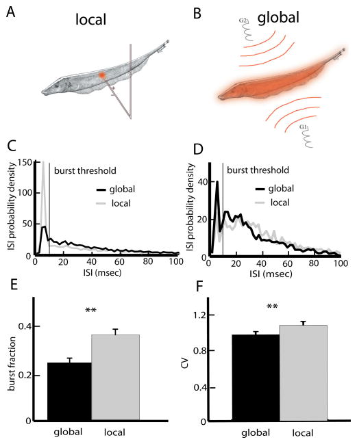
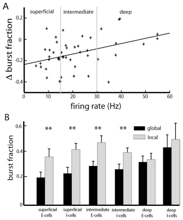
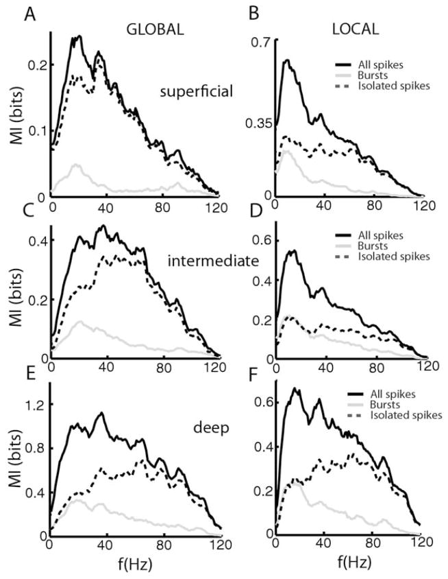
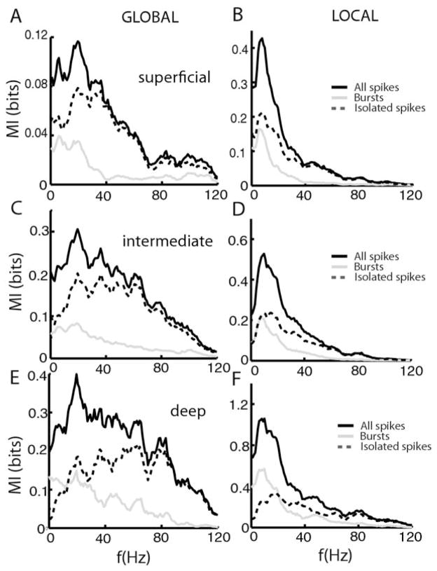
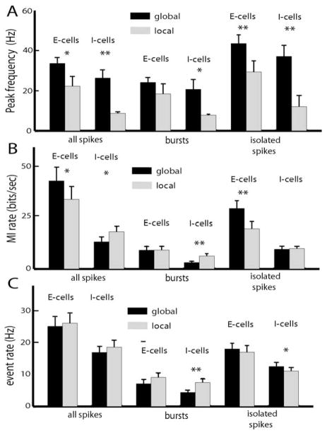
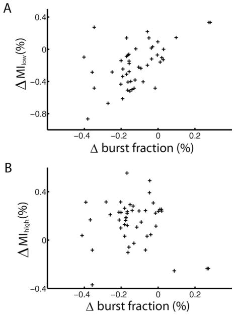
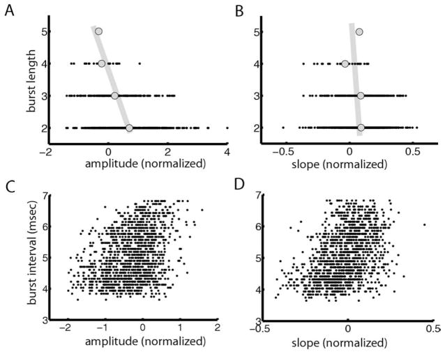
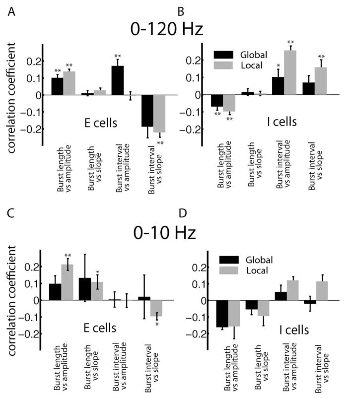
Many neurons tend to fire clusters of action potentials called bursts followed by quiescence in response to sensory input. While the mechanisms that underlie burst firing are generally well understood in vitro, the functional role of these bursts in generating behavioral responses to sensory input in vivo are less clear. Pyramidal cells within the electrosensory lateral line lobe (ELL) of weakly electric fish offer an attractive model system for studying the coding properties of burst firing, because the anatomy and physiology of the electrosensory circuitry are well understood, and the burst mechanism of ELL pyramidal cells has been thoroughly characterized in vitro. We investigated the coding properties of bursts generated by these cells in vivo in response to mimics of behaviorally relevant sensory input. We found that heterogeneities within the pyramidal cell population had quantitative but not qualitative effects on burst coding for the low frequency components of broadband time varying input. Moreover, spatially localized stimuli mimicking, for example, prey tended to elicit more bursts than spatially global stimuli mimicking conspecific-related stimuli. We also found small but significant correlations between burst attributes such as the number of spikes per burst or the interspike interval during the burst and stimulus attributes such as stimulus amplitude or slope. These correlations were much weaker in magnitude than those observed in vitro. More surprisingly, our results show that correlations between burst and stimulus attributes actually decreased in magnitude when we used low frequency stimuli that are expected to promote burst firing. We propose that this discrepancy is attributable to differences between ELL pyramidal cell burst firing under in vivo and in vitro conditions.


Figures

Fig. 1
Fig. 1
ELL pyramidal cells display differential responses to stimuli with differing spatial extents. (A) Local stimulation geometry: a small dipole produces spatially localized AMs of the fish’s own EOD. (B) Global stimulation geometry: two electrodes (G1, G2) located lateral to the animal give rise to spatially diffuse AMs of the fish’s own EOD. (C) ISI probability densities from a representative superficial pyramidal cell under local and global noise stimulation. The noise’s temporal profile was identical in both situations. This cell had a greater tendency to display ISIs less than the burst threshold under local stimulation. (D) The ISI probability densities from an example deep pyramidal cell under local and global noise stimulation were quite similar. (E) Population-averaged burst fractions (i.e. the fraction of ISIs less than the burst threshold) under local and global stimulation. (F) Population averaged ISI coefficient of variation (CV) under local and global stimulation. Asterisks indicate statistical significance at the P=0.05 level using a Signrank test. For interpretation of the references to color in this figure legend, the reader is referred to the Web version of this article.
Fig. 2
Fig. 2
Effects of pyramidal cell heterogeneities on burst firing under local and global stimulation. (A) Change in burst fraction (global-local) as a function of the cell’s baseline firing rate. Superficial pyramidal cells (i.e. cells whose firing rates are less than 15 Hz) display reduced burst fraction under global stimulation but deep pyramidal cells (i.e. cells whose firing rates are greater than 30 Hz) showed little change. (B) Population-averaged burst fractions for E and I-type pyramidal cells of each class. “**” indicates statistical significance with P<0.01 (see text for details).
Fig. 3
Fig. 3
Bursts and Isolated spikes code for different stimulus attributes under local and global stimulation for E-cells. Population-averaged mutual information rate densities for all spikes (black), bursts (gray), and isolated spikes (dashed) for superficial E-cells under global stimulation (A), for superficial E-cells under local stimulation (B), for intermediate E-cells under global stimulation (C), for intermediate E-cells under local stimulation (D), for deep E-cells under global stimulation (E), and for deep E-cells under local stimulation (F).
Fig. 4
Fig. 4
Bursts and Isolated spikes code for different stimulus attributes under local and global stimulation for I-cells. Population averaged mutual information rate densities for all spikes (black), bursts (gray), and isolated spikes (dashed) for superficial I-cells under global stimulation (A), for superficial I-cells under local stimulation (B), for intermediate I-cells under global stimulation (C), for intermediate I-cells under local stimulation (D), for deep I-cells under global stimulation (E), and for deep I-cells under local stimulation (F).
Fig. 5
Fig. 5
Summary of changes in pyramidal cell frequency tuning under local and global stimulation. (A) Population-averaged peak frequency tuning as measured by the mutual information rate density curves for E and I-type pyramidal cells of all three classes. (B) Population-averaged mutual information rates for all spikes, bursts, and isolated spikes obtained for E and I-type pyramidal cells of all three classes. (C) Population-averaged firing rate, burst rate, and isolated spike rate for E and I-type pyramidal cells of all classes. “*” indicates statistical significance with P<0.05 and “**” indicates statistical significance with P<0.01 (see text for details).
Fig. 6
Fig. 6
Correlating changes in burst firing to changes in frequency tuning. (A) The change in low frequency (0–40 Hz) mutual information rate (local-global) plotted as a function of the cell’s change in burst fraction (local-global) showed a significant correlation (R=0.3035, P=0.01, n=47). (B) The change in high frequency (40–80 Hz) mutual information rate (local-global) as a function of the change in burst fraction (local-global) showed no significant correlation (R=0.1012, P=0.4983, n=47).
Fig. 7
Fig. 7
Correlating burst attributes to stimulus attributes for a representative I-cell. (A) Number of spikes per burst (burst length) as a function of stimulus amplitude for an example cell showing a weak but significant negative correlation (R=−0.3367, P≪10−3). (B) Burst length as a function of stimulus slope showing a weak but significant correlation (R=−0.0934, P=0.0054). (C) Burst interval as a function of stimulus amplitude showing a significant correlation (R=0.3611, P≪10−3). (D) Average interspike interval during a burst as a function of stimulus slope showing a significant positive correlation (R=0.4239, P≪10−3).
Fig. 8
Fig. 8
Summary of population-averaged correlation coefficients obtained between burst and stimulus attributes for 0–120 Hz stimuli for E (A) and I (B) cells. The correlation coefficients obtained for 0–10 Hz stimuli for E (C) and I (D) cells are also shown. “**” and “*” indicate statistical significance at the P=0.01 and 0.05 levels using a signrank test, respectively.

Similar articles

Cited by

References

    1. Bannister NJ, Larkman AU. Dendritic morphology of CA1 pyramidal neurones from the rat hippocampus: I. branching patterns. J Comp Neurol. 1995a;360:150–160. - PubMed
    1. Bannister NJ, Larkman AU. Dendritic morphology of CA1 pyramidal neurones from the rat hippocampus: II. spine distributions. J Comp Neurol. 1995b;360:161–171. - PubMed
    1. Bastian J. Gain control in the electrosensory system mediated by descending inputs to the electrosensory lateral line lobe. J Neurosci. 1986;6:553–562. - PMC - PubMed
    1. Bastian J, Chacron MJ, Maler L. Receptive field organization determines pyramidal cell stimulus-encoding capability and spatial stimulus selectivity. J Neurosci. 2002;22:4577–4590. - PMC - PubMed
    1. Bastian J, Chacron MJ, Maler L. Plastic and non-plastic cells perform unique roles in a network capable of adaptive redundancy reduction. Neuron. 2004;41:767–779. - PubMed

Publication types