

Differential regulation of epidermal function by VDR coactivators
- PMID: 20298785
- PMCID: PMC2906691
- DOI: 10.1016/j.jsbmb.2010.03.027
Differential regulation of epidermal function by VDR coactivators
Abstract
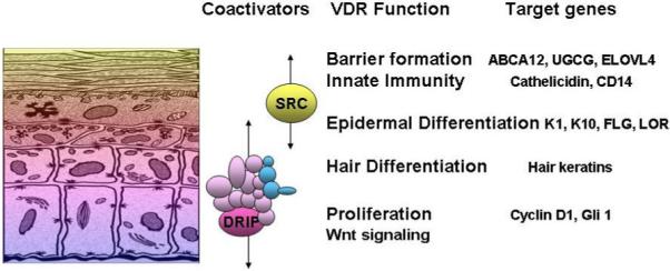
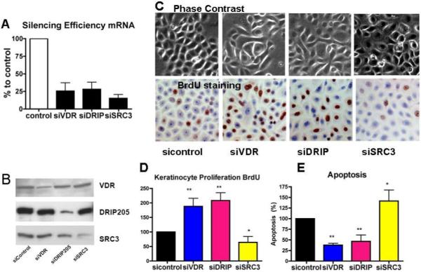
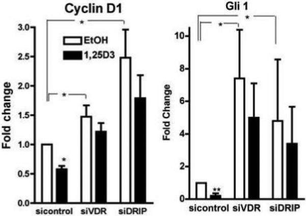
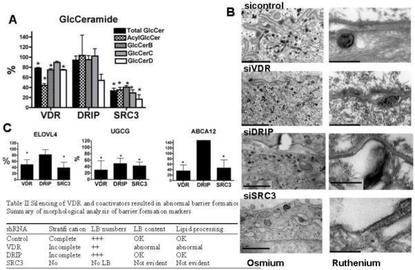
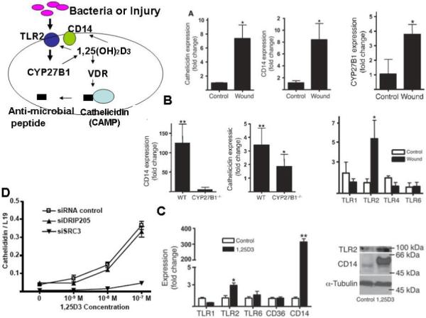
The transcriptional activity of the vitamin D receptor (VDR) is regulated by a number of coactivator and corepressor complexes, which bind to the VDR in a ligand (1,25(OH)2D3) dependent (coactivators) or inhibited (corepressors) process. In the keratinocyte the major coactivator complexes include the vitamin D interacting protein (DRIP) complex and the steroid receptor coactivator (SRC) complexes. These coactivator complexes are not interchangeable in their regulation of keratinocyte proliferation and differentiation. We found that the DRIP complex is the main complex binding to VDR in the proliferating keratinocyte, whereas SRC2 and 3 and their associated proteins are the major coactivators binding to VDR in the differentiated keratinocyte. Moreover, we have found a specific role for DRIP205 in the regulation of beta-catenin pathways regulating keratinocyte proliferation, whereas SRC3 uniquely regulates the ability of 1,25(OH)2D3 to induce more differentiated functions such as lipid synthesis and processing required for permeability barrier formation and the innate immune response triggered by disruption of the barrier. These findings provide a basis by which we can understand how one receptor (VDR) and one ligand (1,25(OH)2D3) can regulate a large number of genes in a sequential and differentiation specific fashion.
Copyright (c) 2010 Elsevier Ltd. All rights reserved.
Figures
Similar articles
-
Differential role of two VDR coactivators, DRIP205 and SRC-3, in keratinocyte proliferation and differentiation.J Steroid Biochem Mol Biol. 2007 Mar;103(3-5):776-80. doi: 10.1016/j.jsbmb.2006.12.069. Epub 2007 Jan 16. J Steroid Biochem Mol Biol. 2007. PMID: 17223341
-
Two distinct coactivators, DRIP/mediator and SRC/p160, are differentially involved in vitamin D receptor transactivation during keratinocyte differentiation.Mol Endocrinol. 2003 Nov;17(11):2329-39. doi: 10.1210/me.2003-0063. Epub 2003 Jul 31. Mol Endocrinol. 2003. PMID: 12893881
-
Vitamin D receptor and coactivators SRC2 and 3 regulate epidermis-specific sphingolipid production and permeability barrier formation.J Invest Dermatol. 2009 Jun;129(6):1367-78. doi: 10.1038/jid.2008.380. Epub 2008 Dec 4. J Invest Dermatol. 2009. PMID: 19052561 Free PMC article.
-
Reciprocal role of vitamin D receptor on β-catenin regulated keratinocyte proliferation and differentiation.J Steroid Biochem Mol Biol. 2014 Oct;144 Pt A:237-41. doi: 10.1016/j.jsbmb.2013.11.002. Epub 2013 Nov 12. J Steroid Biochem Mol Biol. 2014. PMID: 24239508 Free PMC article. Review.
-
Novel mechanisms for the vitamin D receptor (VDR) in the skin and in skin cancer.J Steroid Biochem Mol Biol. 2015 Apr;148:47-51. doi: 10.1016/j.jsbmb.2014.10.017. Epub 2014 Oct 31. J Steroid Biochem Mol Biol. 2015. PMID: 25445917 Free PMC article. Review.
Cited by
-
Lymphoid enhancer-binding factor-1 (LEF1) interacts with the DNA-binding domain of the vitamin D receptor.J Biol Chem. 2011 May 27;286(21):18444-51. doi: 10.1074/jbc.M110.188219. Epub 2011 Apr 6. J Biol Chem. 2011. PMID: 21471213 Free PMC article.
-
The retinoid X receptors and their ligands.Biochim Biophys Acta. 2012 Jan;1821(1):21-56. doi: 10.1016/j.bbalip.2011.09.014. Epub 2011 Oct 1. Biochim Biophys Acta. 2012. PMID: 22020178 Free PMC article. Review.
-
Expression of epidermal CAMP changes in parallel with permeability barrier status.J Invest Dermatol. 2011 Nov;131(11):2263-70. doi: 10.1038/jid.2011.210. Epub 2011 Jul 28. J Invest Dermatol. 2011. PMID: 21796152 Free PMC article.
-
Halting the March: Primary Prevention of Atopic Dermatitis and Food Allergies.J Allergy Clin Immunol Pract. 2020 Mar;8(3):860-875. doi: 10.1016/j.jaip.2019.12.005. J Allergy Clin Immunol Pract. 2020. PMID: 32147139 Free PMC article.
-
Role of micronutrients in skin health and function.Biomol Ther (Seoul). 2015 May;23(3):207-17. doi: 10.4062/biomolther.2015.003. Epub 2015 May 1. Biomol Ther (Seoul). 2015. PMID: 25995818 Free PMC article. Review.
References
-
- Bikle DD, Pillai S. Vitamin D, calcium, and epidermal differentiation. Endocrine Review. 1993;14:3–19. - PubMed
-
- Pillai S, Bikle DD. Role of intracellular-free calcium in the cornified envelope formation of keratinocytes: differences in the mode of action of extracellular calcium and 1,25 dihydroxyvitamin D3. J Cell Physiol. 1991;146(1):94–100. - PubMed
-
- Bikle DD, Pillai S, Gee E. Squamous carcinoma cell lines produce 1,25 dihydroxyvitamin D, but fail to respond to its prodifferentiating effect. J Invest Dermatol. 1991;97(3):435–441. - PubMed
-
- Hosomi J, Hosoi J, Abe E, et al. Regulation of terminal differentiation of cultured mouse epidermal cells by 1 alpha,25-dihydroxyvitamin D3. Endocrinology. 1983;113(6):1950–1957. - PubMed
-
- Smith EL, Walworth NC, Holick MF. Effect of 1 alpha,25-dihydroxyvitamin D3 on the morphologic and biochemical differentiation of cultured human epidermal keratinocytes grown in serum-free conditions. J Invest Dermatol. 1986;86(6):709–714. - PubMed
Publication types
MeSH terms
Substances
Grants and funding
LinkOut - more resources
Full Text Sources
Other Literature Sources
Miscellaneous
